Comparative Study
. 2010 Feb 23;107(8):3570-5.
doi: 10.1073/pnas.0906596107. Epub 2010 Feb 5.

Genomic code for Sox10 activation reveals a key regulatory enhancer for cranial neural crest

Affiliations
Comparative Study

Genomic code for Sox10 activation reveals a key regulatory enhancer for cranial neural crest

Paola Betancur et al. Proc Natl Acad Sci U S A. .

Abstract

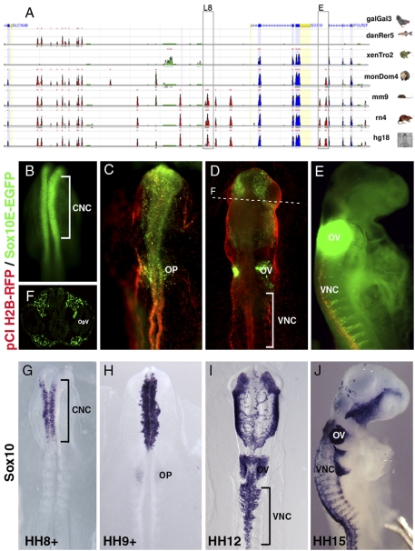
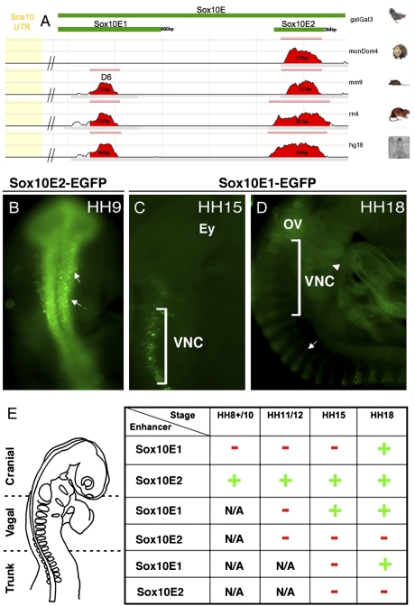
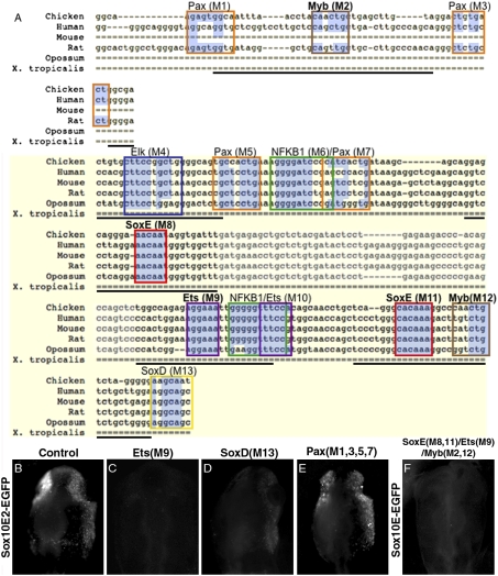
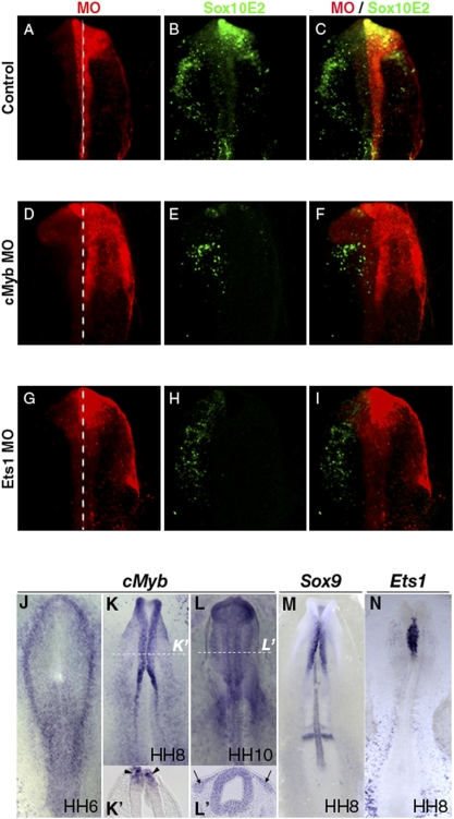
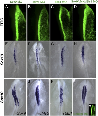
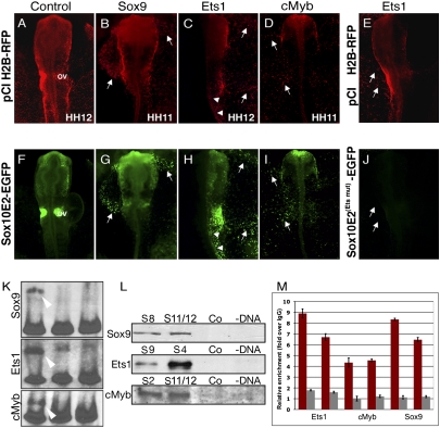
The neural crest is a multipotent, stem cell-like population that migrates extensively in the embryo and forms a wide array of derivatives, ranging from neurons to melanocytes and cartilage. Analyses of the gene regulatory network driving neural crest development revealed Sox10 as one of the earliest neural crest-specifying genes, cell-autonomously driving delamination and directly regulating numerous downstream effectors and differentiation gene batteries. In search of direct inputs to the neural crest specifier module, we dissected the chick Sox10 genomic region and isolated two downstream regulatory regions with distinct spatiotemporal activity. A unique element, Sox10E2 represents the earliest-acting neural crest cis-regulatory element, critical for initiating Sox10 expression in newly formed cranial, but not vagal and trunk neural crest. A second element, Sox10E1, acts in later migrating vagal and trunk crest cells. Deep characterization of Sox10E2 reveals Sox9, Ets1, and cMyb as direct inputs mediating enhancer activity. ChIP, DNA-pull down, and gel-shift assays demonstrate their direct binding to the Sox10E2 enhancer in vivo, whereas mutation of their corresponding binding sites, or inactivation of the three upstream regulators, abolishes both reporter and endogenous Sox10 expression. Using cis-regulatory analysis as a tool, our study makes critical connections within the neural crest gene regulatory network, thus being unique in establishing a direct link of upstream effectors to a key neural crest specifier.


Conflict of interest statement

The authors declare no conflict of interest.

Figures

Fig. 1.
Fig. 1.
Sox10 cis-regulatory analysis. (A) Schematic diagram showing comparative genomic analysis using the ECR browser. Chicken, zebrafish, Xenopus, opossum, mouse, rat, and human genomic sequences were compared between Sox10 and neighboring genes, Slc16A8 and PolR2F: (red) highly conserved elements; (blue) coding exons; (green) transposable elements and simple repeats. Boxed Sox10 putative regulatory regions L8 (late) and E (early) show activity in neural crest. UTRs shaded in yellow. (B) At HH8+, GFP transcripts are detected by fluorescent in situ hybridization in CNC, similar to endogenous Sox10 expression (G). Distribution of EGFP transcripts (C–E) (HH9+, HH12, HH15) is similar to endogenous Sox10 in H to J, respectively. (D) EGFP expression at HH12 in rhombomere5 stream surrounding otic vesicle (OV) resembles endogenous Sox10 (I), but is missing in vagal neural crest (VNC). (F) Cross section of embryo in D shows specific Sox10E regulatory activity in CNC around optic vesicle (OpV). (G–J) Endogenous Sox10 expression at HH8 ± 15. OP, otic placode.
Fig. 2.
Fig. 2.
Sox10E contains distinct cranial and vagal/trunk regulatory elements. (A) Schematic diagram representing dissection of Sox10E fragment, located ∼1-kb downstream of Sox10 locus (UTR in yellow). Two smaller active regulatory fragments embedded within Sox10E, Sox10E1, and E2, each contain a conserved region (red bar) with 70% sequence homology between amniotes. (B) Sox10E2 drives expression in delaminating CNC (arrows) at HH8+; (C) Sox10E1 is first active in migrating VNC at HH15. (D) Sox10E1 activity persists in migrating VNC, trunk neural crest (arrow), and branchial arches 3 to 5 (arrowhead). (E) Table S1 summarizes distinct temporal (HH9–18) and spatial (cranial/vagal/trunk) regulatory activity of Sox10E1 and E2. (Red −) no expression; (green +) EGFP reporter expression.
Fig. 3.
Fig. 3.
Sox10E2 transcriptional inputs. (A) Schematic diagram showing sequence alignment of 264-bp Sox10E2 region; essential core region shaded in yellow. Colored frames indicate computationally identified putative transcription factors binding motifs. Mutations M1 to M13 were replaced by random sequences. Faded sequence shows a 45-bp region deleted or replaced by mCherry coding sequence. Highlighted in blue are conserved nucleotides within putative binding motifs. Single dashed lines indicate absent bases in the alignment and thick dashed lines nonalignable sequences. Thick solid underlines delineate Sox10E2 subfragments used in EMSA and pull-down assays. Sox10E2-driven EGFP expression in CNC (B) is abolished upon mutation of an Ets1 binding motif (C), but only decreased after mutation of putative SoxD motif (D), and not affected by simultaneous mutation of four putative Pax sites (E). (F) Simultaneous inactivation of SoxE, Ets, and Myb binding sites (M2, M8, M9, M11, M12) within a large genomic region abolishes reporter expression in delaminating CNC.
Fig. 4.
Fig. 4.
Ets1 and cMyb are necessary for activation of Sox10E2 element. (A) Control morpholino (Right; red) has no effect on Sox10E2-driven Cherry (B) (green) compared to nonelectroporated (Left) side. cMyb MO (D) significantly reduces, whereas Ets1 MO (G) abolishes Sox10E2-driven Cherry expression, (E and H, respectively; C, F, and I are merged images of A/B, D/E, and G/H, respectively). White dotted lines indicate the midline. Green/red channels are inverted for consistency. (J–L) Endogenous cMyb, Sox9, and Ets1 expression precedes that of Sox10. At HH6, cMyb is expressed within the neural plate border (J) and confined to dorsal neural folds by HH8 (K and K, arrowheads). At HH10, cMyb is observed in migrating neural crest (L and section at dotted line, Larrows). At HH8, before Sox10 onset, Sox9 (M) and Ets1 (N) are expressed by presumptive CNC in the dorsal neural tube.
Fig. 5.
Fig. 5.
Sox9, cMyb, and Ets1 are required for endogenous Sox10 expression in delaminating CNC cells. HH8+ embryos with unilateral electroporation of Sox9 (A and E), cMyb (B and F), and Ets1 (C and G), but not of control (L and M) morpholino (MO) (green) show significant decrease in endogenous Sox10 expression in delaminating CNC compared with nonelectroporated side. Coelectroporation of Sox9, cMyb, Ets1 MOs completely abolish endogenous Sox10 expression (D and H). Showing specificity, the effect is rescued by morpholino coelectroporation with corresponding expression construct (I, J, and K). (K). Statistical relevance by χ2 test of MOs on Sox10 expression was P < 0.02; of rescues was P < 0.03 (Sox9; Ets1) and P ≤ 0.04 (cMyb).
Fig. 6.
Fig. 6.
Overexpression of Sox9 (B and G), Ets1 (C and H), or cMyb (D and I), but not of control plasmid, pCI H2B-RFP (A and F), ectopically activates Sox10E2-driven EGFP expression in extra-embryonic ectoderm (white arrows). In H, arrowheads show ectopic expression in posterior neural tube. Misexpression of Ets1 (E) fails to activate ectopic EGFP expression (J, arrows) in mutated Sox10E2 construct lacking an Ets binding motif (M9). (K) EMSA shows a clear band shift (white arrowhead) when nuclear extracts containing overexpressed Sox9, Ets1, or cMyb proteins are combined with Sox10E2 subfragments, S11, S9, and S2, respectively (Lane 1). This binding is outcompeted when excess nonlabeled probe is added (Lane 2) and absent from nuclear extracts from control plasmid-transfected cells (Lane 3). (L) Biotinylated Sox10E2 subfragments (S8, S11-Sox9, S4, S9-Ets1, and S2, S12-cMyb), as well as scrambled control fragments and noncoated Dynal streptavidin beads, used as bait in a DNA pull-down assay, show specific transcription factor binding as analyzed on a Western blot. (M) Direct binding of Ets1, cMyb, and Sox9 to the Sox10E2 enhancer element in vivo as assessed by qChIP. Binding to Sox10E2 (red bars) or control region (gray bars) was assessed with two primer sets for each region and expressed as relative enrichment of target over control antibody; graph reflects mean ± SD from a representative experiment. qChIP was performed three to four times for each factor. Enrichment relative to input DNA from all independent experiments is presented in Fig. S8.

Similar articles

Cited by

References

    1. Martinsen BJ, Bronner-Fraser M. Neural crest specification regulated by the helix-loop-helix repressor Id2. Science. 1998;281:988–991. - PubMed
    1. Tahtakran SA, Selleck MA. Ets-1 expression is associated with cranial neural crest migration and vasculogenesis in the chick embryo. Gene Expr Patterns. 2003;3:455–458. - PubMed
    1. Meulemans D, Bronner-Fraser M. Gene-regulatory interactions in neural crest evolution and development. Dev Cell. 2004;7:291–299. - PubMed
    1. Nikitina N, Sauka-Spengler T, Bronner-Fraser M. Dissecting early regulatory relationships in the lamprey neural crest gene network. Proc Natl Acad Sci USA. 2008;105:20083–20088. - PMC - PubMed
    1. Sauka-Spengler T, Bronner-Fraser M. Development and evolution of the migratory neural crest: a gene regulatory perspective. Curr Opin Genet Dev. 2006;16:360–366. - PubMed

Publication types

Substances