. 2010 Apr;76(7):2271-9.
doi: 10.1128/AEM.02361-09. Epub 2010 Feb 5.

Role of extracellular DNA during biofilm formation by Listeria monocytogenes

Affiliations

Role of extracellular DNA during biofilm formation by Listeria monocytogenes

Morten Harmsen et al. Appl Environ Microbiol. 2010 Apr.

Abstract

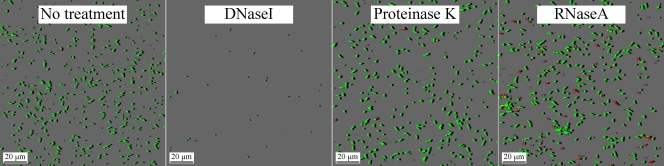
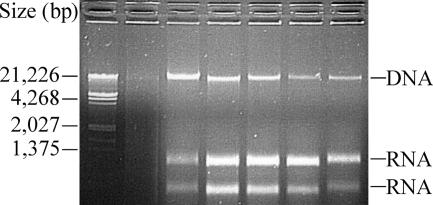
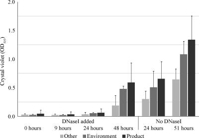
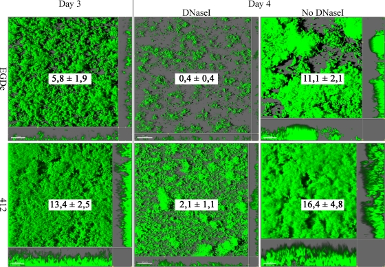
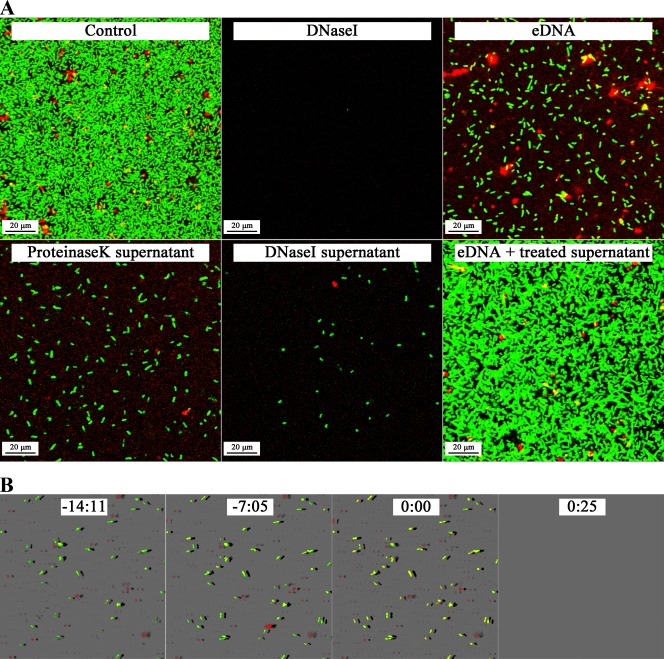
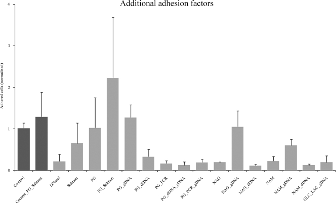
Listeria monocytogenes is a food-borne pathogen that is capable of living in harsh environments. It is believed to do this by forming biofilms, which are surface-associated multicellular structures encased in a self-produced matrix. In this paper we show that in L. monocytogenes extracellular DNA (eDNA) may be the only central component of the biofilm matrix and that it is necessary for both initial attachment and early biofilm formation for 41 L. monocytogenes strains that were tested. DNase I treatment resulted in dispersal of biofilms, not only in microtiter tray assays but also in flow cell biofilm assays. However, it was also demonstrated that in a culture without eDNA, neither Listeria genomic DNA nor salmon sperm DNA by itself could restore the capacity to adhere. A search for additional necessary components revealed that peptidoglycan (PG), specifically N-acetylglucosamine (NAG), interacted with the DNA in a manner which restored adhesion. If a short DNA fragment (less than approximately 500 bp long) was added to an eDNA-free culture prior to addition of genomic or salmon sperm DNA, adhesion was prevented, indicating that high-molecular-weight DNA is required for adhesion and that the number of attachment sites on the cell surface can be saturated.


Figures

FIG. 1.
FIG. 1.
L. monocytogenes EGDe attached cells after 30 min of incubation at 37°C in Nunc coverglass cell culture chambers. The images show L. monocytogenes cells adhering in the presence of only PBS buffer (no-treatment control) and in the presence of PBS buffer containing enzymes (DNase I, proteinase K, and RNase A). The cells were stained with SYTO9 and propidium iodide, which resulted in live green cells and dead red cells.
FIG. 2.
FIG. 2.
Agarose (1%, wt/vol) gel electrophoresis of ethanol-precipitated eDNA. The lane on the left contained lambda marker DNA digested with HindIII and EcoRI, the next lane contained nothing, and the remaining lanes contained precipitated DNA from ON cultures of different L. monocytogenes strains. Sizes are indicated on the left.
FIG. 3.
FIG. 3.
Graph showing the results of the microtiter biofilm assay as assessed by crystal violet staining. Forty-one strains (see Table 1) were divided into three major groups based on their origins (product [8 strains], environment [14 strains], and other [19 strains]) (see Table 1). Some cultures were treated with DNase I at specific time points after inoculation (0, 9, 24, and 48 h). Other cultures were used as controls to which DNase I was not added and were examined after 24 and 51 h. Except for the 24-h control to which no DNase I was added, all values were obtained using crystal violet staining at 51 h after inoculation. For the 24-h control to which no DNase I was added the values were obtained using crystal violet staining at 24 h after inoculation. The values are averages and standard deviations. The results are expressed as optical densities at 595 nm. The assay was performed at 37°C.
FIG. 4.
FIG. 4.
Flow cell biofilms of the L. monocytogenes EGDe and 412 strains. The upper panels show biofilm formation by the EGDe strain, and the lower panels show biofilm formation by the 412 strain. Both biofilms were grown in medium containing 2.5% (vol/vol) BHI at 37°C. The cells were tagged with GFP and therefore are green. The large images are three-dimensional top-down images of the biofilms, and the small images to the right of and below the large images are side views of sections. The day 3 images are images of biofilms 72 h after inoculation. The day 4 images are images of DNase I-treated biofilms 96 h after inoculation and after 18 h of DNase I treatment and images a biofilm that was not treated with DNase I 96 h after inoculation. The quantities (means ± standard deviations) of biomass volume divided by substratum area (μm3/μm2) determined by COMSTAT analysis are indicated in each large image. The biomass was quantified by determining the average for 18 images taken in two channels of the flow cell.
FIG. 5.
FIG. 5.
L. monocytogenes EGDe attached cells after 30 min of incubation at 37°C in Nunc coverglass cell culture chambers. All images show attachment of cells in early exponential phase after 30 min of attachment at 37°C. (A) Adhered cells were grown until the OD600 was 0.5 in BHI medium at 37°C with 100 μg ml−1 DNase I (the control was grown without DNase I) prior to attachment, and all cell suspensions were washed in PBS buffer to remove the remaining DNase I. Different supplements were added to determine their effects on attachment, except for the control and DNaseI cultures, which contained only cells grown without DNase I and cells grown with DNase I, respectively. The eDNA culture was supplemented with gel-purified eDNA. The proteinase K and DNase I cultures were supplemented with proteinase K-treated supernatant and DNase I-treated supernatant, respectively. The remaining image (eDNA + treated supernatant) shows cells grown with gel-purified eDNA and supernatant that were treated with proteinase K and DNase I. (B) Time series for adhered cells treated with 100 μg ml−1 DNase I. The time is indicated at the top in each image and is expressed as minutes:seconds. At time point 0 the cells were treated with DNase I; a negative time indicates the time before treatment. The image on the right was obtained 25 s after treatment. The cells were tagged with GFP and therefore are green in the images. SYTOX orange was used as a DNA stain, resulting in the red regions in the images.
FIG. 6.
FIG. 6.
Numbers of cells (means and standard deviations) adhering after 15 min of incubation at 37°C. Cells adhered in flow cell chambers containing HTM with 3% (wt/vol) glucose, and attachment was induced with DNA that differed in origin and length in combination with additional compounds. The adhered cells were counted, and the values were normalized to the average number of cells adhering in the control, which contained exponentially growing cells of the EGDe strain. The control cells and the control cells treated with peptidoglycan and salmon sperm DNA were not treated with DNase I, whereas all other cells were treated with 100 μg ml−1 DNase I for 30 min prior to adhesion. Abbreviations: PG, peptidoglycan; Salmon, salmon sperm DNA; gDNA, Listeria genomic DNA; dDNA, digested Listeria genomic DNA; PCR, 200-bp fragment of Listeria DNA; NAG, N-acetylglucosamine; NAM, N-acetylmuramic acid; GLC, glucose; LAC, lactic acid. DNA and additional compounds were all added at a final concentration of 3 μg ml−1. The cells that were treated with peptidoglycan, digested genomic DNA, and genomic DNA and the cells that were treated with peptidoglycan, the 200-bp fragment of Listeria DNA, and genomic DNA were pretreated with either digested genomic DNA or the 200-bp fragment of Listeria DNA for 5 min prior to addition of the genomic DNA.

Similar articles

Cited by

References

    1. Allesen-Holm, M., K. B. Barken, L. Yang, M. Klausen, J. S. Webb, S. Kjelleberg, S. Molin, M. Givskov, and T. Tolker-Nielsen. 2006. A characterization of DNA release in Pseudomonas aeruginosa cultures and biofilms. Mol. Microbiol. 59:1114-1128. - PubMed
    1. Chae, M. S., and H. Schraft. 2000. Comparative evaluation of adhesion and biofilm formation of different Listeria monocytogenes strains. Int. J. Food Microbiol. 62:103-111. - PubMed
    1. Costerton, J. W., P. S. Stewart, and E. P. Greenberg. 1999. Bacterial biofilms: a common cause of persistent infections. Science 284:1318-1322. - PubMed
    1. Cucarella, C., C. Solano, J. Valle, B. Amorena, I. Lasa, and J. R. Penades. 2001. Bap, a Staphylococcus aureus surface protein involved in biofilm formation. J. Bacteriol. 183:2888-2896. - PMC - PubMed
    1. Dell'Anno, A., and R. Danovaro. 2005. Extracellular DNA plays a key role in deep-sea ecosystem functioning. Science 309:2179. - PubMed

Publication types

LinkOut - more resources