. 2011 Jan;44(1):40-52.
doi: 10.1165/rcmb.2009-0197OC. Epub 2010 Feb 5.

Non-muscle myosin light chain kinase isoform is a viable molecular target in acute inflammatory lung injury

Affiliations

Non-muscle myosin light chain kinase isoform is a viable molecular target in acute inflammatory lung injury

Tamara Mirzapoiazova et al. Am J Respir Cell Mol Biol. 2011 Jan.

Abstract

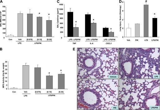
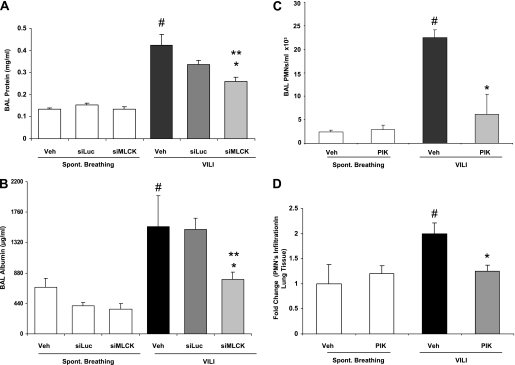
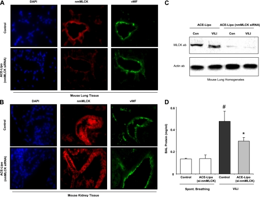
Acute lung injury (ALI) and mechanical ventilator-induced lung injury (VILI), major causes of acute respiratory failure with elevated morbidity and mortality, are characterized by significant pulmonary inflammation and alveolar/vascular barrier dysfunction. Previous studies highlighted the role of the non-muscle myosin light chain kinase isoform (nmMLCK) as an essential element of the inflammatory response, with variants in the MYLK gene that contribute to ALI susceptibility. To define nmMLCK involvement further in acute inflammatory syndromes, we used two murine models of inflammatory lung injury, induced by either an intratracheal administration of lipopolysaccharide (LPS model) or mechanical ventilation with increased tidal volumes (the VILI model). Intravenous delivery of the membrane-permeant MLC kinase peptide inhibitor, PIK, produced a dose-dependent attenuation of both LPS-induced lung inflammation and VILI (~50% reductions in alveolar/vascular permeability and leukocyte influx). Intravenous injections of nmMLCK silencing RNA, either directly or as cargo within angiotensin-converting enzyme (ACE) antibody-conjugated liposomes (to target the pulmonary vasculature selectively), decreased nmMLCK lung expression (∼70% reduction) and significantly attenuated LPS-induced and VILI-induced lung inflammation (∼40% reduction in bronchoalveolar lavage protein). Compared with wild-type mice, nmMLCK knockout mice were significantly protected from VILI, with significant reductions in VILI-induced gene expression in biological pathways such as nrf2-mediated oxidative stress, coagulation, p53-signaling, leukocyte extravasation, and IL-6-signaling. These studies validate nmMLCK as an attractive target for ameliorating the adverse effects of dysregulated lung inflammation.


Figures

Figure 1.
Figure 1.
PIK attenuates LPS-induced vascular permeability and lung injury in vitro and in vivo. (A) Human lung endothelia were plated on gold microelectrodes, cultured to confluence, and challenged with agonist, as indicated. Measurements of TER were recorded, and the data depicted are representative of three independent experiments. Shown are the responses to LPS (100 ng/ml), PIK (250 μM), or a combination of the two agents. Lipopolysaccharide elicits significant and sustained decreases in TER. PIK alone caused a prolonged, significant initial increase in TER, and restored TER values in LPS-challenged cells. Inset: Example of MLC phosphorylation after PIK/LPS treatment. Veh, vehicle. (B) Dose-dependent inhibition of MLC phosphorylation by PIK after LPS challenge. The phosphorylation of MLC was detected in mouse lungs after LPS/PIK treatment, as described in Materials and Methods. Inset: PIK alone (0.25 mg/mouse) did not alter basal levels of MLC phosphorylation. Representative Western blot with quantification shows dose-dependent decreases of MLC phosphorylation (0.075–0.25 mg PIK per mouse) compared with LPS. Values are presented as ratios of densitometry units of ppMLC to total MLC. (C and D) Assessments of BAL albumin and tissue albumin concentrations after LPS/PIK. Lipopolysaccharide induced increases in albumin leakage from the vascular space into surrounding lung tissues and BAL, which were significantly reduced in PIK-treated mice. *Significant differences between LPS and LPS/PIK groups. #Significant differences in vehicle versus LPS-challenged mice. Overall, the concentration of albumin in BAL fluids was reduced in a dose-dependent manner by PIK.
Figure 2.
Figure 2.
PIK reduces LPS-induced accumulation of WBCs in BAL. (A) LPS-mediated cell infiltration into lung tissue was reduced in a dose-dependent manner by intravenous administration of PIK. *Significant differences in LPS versus LPS/PIK . Con, control; Veh, vehicle. (B) Activities of MPO were elevated in lung tissue from LPS/vehicle groups, but decreased in LPS/PIK-treated mice (0.15 PIK/LPS versus LPS, *P < 0.01; 0.25 PIK/LPS versus LPS, *P < 0.01). (C) Effects of PIK on BAL cytokine levels in C57BL/6 mice after challenge with LPS . Concentrations of TNF-α, IL-6, and CXCL1 in BAL were significantly reduced compared with LPS alone. *Significant reduction resulting from PIK. (D) Histologic assessment of PIK's effects on LPS-induced lung inflammation. Lungs were analyzed for interstitial edema and inflammatory cell infiltration. Paraffin-embedded hematoxylin-and-eosin–stained sections were prepared by standard techniques. Sagittal sections through central and peripheral areas of each lobe were obtained, to represent histologic changes in the entire lung (n = 3 from each treatment group). Quantitative analyses of lung inflammation, using parameters of severity (score, 0–3), focal/diffuse inflammation (score, 0–3), and parenchyma/airway inflammation (score, 0–3), demonstrated fewer pathologic changes in LPS-challenged mice receiving PIK. *Significant difference produced by PIK. #Significant difference produced by LPS compared with vehicle. PIK alone did not induce inflammation. (E ) Hematoxylin-and-eosin staining of lung sections indicated that LPS-induced edema and leukocyte infiltration were reversed by administration of PIK (0.25 mg/mouse).
Figure 3.
Figure 3.
(A) Endothelial cells were cultured to confluence on gold microelectrodes and exposed to LPS (100 ng/ml), as indicated. Transendothelial electrical resistance was recorded for 18 hours of PIK (250 μM), administered 3 hours after LPS challenge, resulting in an increase of TER to initial basal level in LPS-challenged cells. (B) Lipopolysaccharide-mediated protein accumulation into mouse BAL fluid was reduced by intravenous injection of PIK (0.25 mg/mouse), 3 hours after LPS challenge. *Significant difference. (C) The BAL neutrophil count was sufficiently decreased in the LPS/PIK group, compared with LPS alone. *Significant difference.
Figure 4.
Figure 4.
In vitro and in vivo silencing of nmMLCK expression attenuated LPS-induced endothelial barrier dysfunction. (A) Endothelial cells were cultured to confluence on gold microelectrodes and exposed to LPS (100 ng/ml) as indicated, with transendothelial electrical resistance (TER) recorded for 20 hours. Lipopolysaccharide elicited a significant, sustained decease in TER, with the nmMLCK siRNA abolishing the disruptive effect of LPS. Data are expressed as mean ± SE. (B) The siRNA specific for mouse nmMLCK was directly injected as described in Materials and Methods. Western blots of nmMLCK from mouse lung lysates show decreased nmMLCK expression (∼80%) between the control/LPS group and the siRNA/LPS group, 72 hours after silencing. Inset: The decreased expression of nmMLCK in ECs after treatment with siRNA. *Significant difference between control group and siMLCK or siMLCK/LPS group. #Significant difference between LPS group and siMLCK or siMLCK/LPS group. The β-actin blot confirms equal protein loading. The blot shown is representative of at least of three experiments. (C) Mouse lungs with combined siMLCK/LPS challenge produced evidence for decreased epithelial–endothelial permeability, as reflected by BAL protein concentrations, whereas LPS or siLuc/LPS treatment exhibited significantly increased BAL protein. Lipopolysaccharide-induced elevation of BAL protein was significantly reduced by nmMLCK siRNA. *,#Significant decreases compared with LPS or siLuc/LPS. (D) The increase in number of BAL neutrophils (PMNs) in LPS-challenged mice. The BAL neutrophil counts were significantly decreased in the siMLCK/LPS group, compared with LPS or LPS/siLuc mice. *,#Significant decreases (P < 0.05).
Figure 5.
Figure 5.
Effects of PIK and nmMLCK silencing on murine VILI. Veh, vehicle; Spont. Breathing, spontaneously breathing. (A) The concentration of BAL protein was significantly elevated in all VILI-treated groups compared with SB groups. In addition, when mice were treated with nmMLCK siRNA and exposed to VILI, the siMLCK/VILI group had a significantly lower concentration of BAL protein. *,**,#Significant differences. (B) Evaluation of BAL albumin concentration showed that mice receiving nmMLCK siRNA had significantly lower concentrations of albumin compared with mice exposed only to mechanical ventilation with vehicle or control siRNA. *,**,#Significant differences. (C) Increased BAL neutrophil invasion, associated with VILI, was significantly attenuated after administration of PIK. *Significant difference between VILI and VILI/PIK group. #Significant difference between VILI versus vehicle or PIK alone. (D) Administration of PIK significantly reduced the fold change in neutrophil migration in lung tissue. *Significant difference between the VILI and VILI/PIK groups. #Significant difference between VILI versus vehicle or PIK alone.
Figure 6.
Figure 6.
Attenuation of VILI by intravenously administered ACE antibody-conjugated liposomes (ACE-Lipo), with nmMLCK siRNA as cargo. (A) Immunofluorescence analysis of lung tissue (in vivo targeting of microvascular endothelial cells). The ACE antibody (ab)-conjugated liposomes (containing siRNA against nmMLCK) were injected into the jugular vein, to deliver nmMLCK siRNA specifically to lung vessels. After 5 days, tissues were assessed by examining sections under a fluorescence microscope, revealing that pulmonary vessels from mice treated with ACE antibody-conjugated liposomes exhibited reduced nmMLCK expression (red), compared with vessels from control mice. The vascular lumen is identified by vessel staining with endothelial cell-specific von Willebrand Factor antibody (green). (B) Immunohistochemical analysis of mouse kidney tissues from mice challenged with ACE antibody-conjugated liposomes (containing siRNA against nmMLCK) indicates no changes in nmMLCK expression within the renal vasculature, compared with control mice. (C) Liposomal delivery of nmMLCK siRNA decreases nmMLCK expression in mouse lung homogenates. A representative Western blot is shown (representing at least three experiments). (D) Graphic representation of BAL protein indicates that ACE antibody-conjugated liposome delivery significantly reduces VILI-induced lung injury. *,#Significant differences (P < 0.05). Spont. Breathing, spontaneously breathing.
Figure 7.
Figure 7.
Deletion of nmMLCK gene (nmMLCK−/−) results in protection from severe murine ventilator-induced lung injury. Spont. Breathing, spontaneously breathing. (A) In contrast to increase in BAL protein induced by VILI in WT mice, this effect was largely abrogated in nmMLCK−/− mice (n = 6 animals per group). (B) Ventilator-induced lung injury induces increased lung microvascular permeability, as reflected by BAL albumin, compared with SB mice. In addition, nmMLCK−/− mice are less susceptible to VILI, and exhibit significantly reduced BAL albumin concentrations, compared with WT mice exposed to VILI. (C) Gene expression pattern of differentially expressed genes in VILI-challenged WT mice and nmMLCK KO mice. Gene identity is shown at the right of each row. The overlap of Gene Lists 1 and 3 was classified by dChip (http://biosun1.harvard.edu/complab/dchip/). Red, white, and blue indicate expression above, at, and below mean level, respectively.
Figure 8.
Figure 8.
Ingenuity Pathways Analysis of VILI-dysregulated genes in WT and nmMLCK−/− mice. (A) Significant canonical pathways were enriched with dysregulated genes in WT-VILI (blue) and KO-VILI animals (red). (B) Fold changes of dysregulated genes, induced by WT–VILI or KO–VILI in the leukocyte extravasation signaling pathway. All genes shown were identified by SAM software, using criteria of fold change > 3 and false discovery rate < 1% (Table E1). (C) Bleeding times (in seconds) were analyzed in genetically engineered nmMLCK KO and WT mice. The nmMLCK−/− mice exhibited significantly prolonged bleeding times (1.5-fold longer).

References

    1. Lazar V, Garcia JG. A single human myosin light chain kinase gene (MLCK; MYLK) transcribes multiple nonmuscle isoforms. Genomics 1999;57:256–267. - PubMed
    1. Dreyfuss D, Saumon G. Ventilator-induced lung injury: lessons from experimental studies. Am J Respir Crit Care Med 1998;157:294–323. - PubMed
    1. Ma SF, Grigoryev DN, Taylor AD, Nonas S, Sammani S, Ye SQ, Garcia JG. Bioinformatic identification of novel early stress response genes in rodent models of lung injury. Am J Physiol 2005;289:L468–L477. - PubMed
    1. Chopra M, Reuben JS, Sharma AC. Acute lung injury: apoptosis and signaling mechanisms. Exp Biol Med 2009;234:361–371. - PubMed
    1. Petrache I, Verin AD, Crow MT, Birukova A, Liu F, Garcia JG. Differential effect of MLC kinase in TNF-alpha–induced endothelial cell apoptosis and barrier dysfunction. Am J Physiol 2001;280:L1168–L1178. - PubMed

Publication types

MeSH terms