. 2010 Apr 30;285(18):13966-78.
doi: 10.1074/jbc.M109.061127. Epub 2010 Feb 6.

Trans-synaptic adhesions between netrin-G ligand-3 (NGL-3) and receptor tyrosine phosphatases LAR, protein-tyrosine phosphatase delta (PTPdelta), and PTPsigma via specific domains regulate excitatory synapse formation

Affiliations

Trans-synaptic adhesions between netrin-G ligand-3 (NGL-3) and receptor tyrosine phosphatases LAR, protein-tyrosine phosphatase delta (PTPdelta), and PTPsigma via specific domains regulate excitatory synapse formation

Seok-Kyu Kwon et al. J Biol Chem. .

Abstract

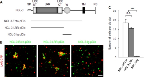
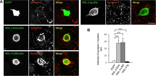
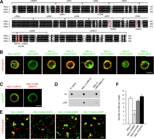
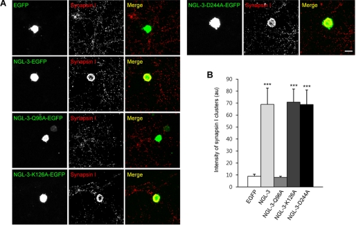
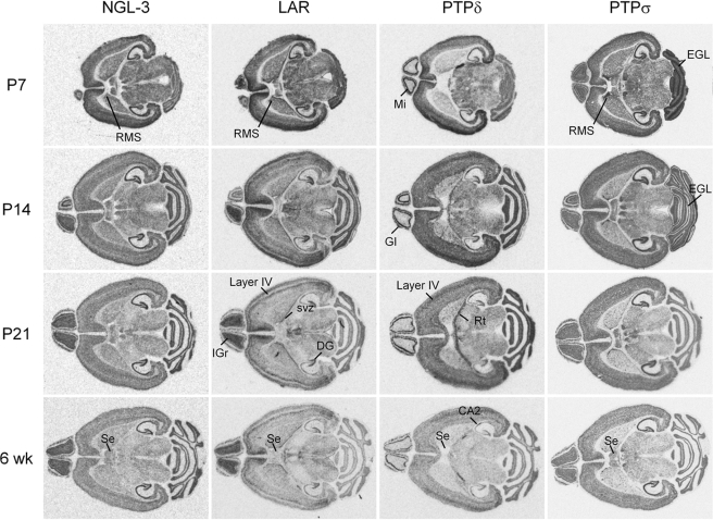
Synaptic cell adhesion molecules regulate various steps of synapse formation. The trans-synaptic adhesion between postsynaptic NGL-3 (for netrin-G ligand-3) and presynaptic LAR (for leukocyte antigen-related) regulates excitatory synapse formation in a bidirectional manner. However, little is known about the molecular details of the NGL-3-LAR adhesion and whether two additional LAR family proteins, protein-tyrosine phosphatase delta (PTPdelta), and PTPsigma, also interact with NGL-3 and are involved in synapse formation. We report here that the leucine-rich repeat (LRR) domain of NGL-3, containing nine LRRs, interacts with the first two fibronectin III (FNIII) domains of LAR to induce bidirectional synapse formation. Moreover, Gln-96 in the first LRR motif of NGL-3 is critical for LAR binding and induction of presynaptic differentiation. PTPdelta and PTPsigma also interact with NGL-3 via their first two FNIII domains. These two interactions promote synapse formation in a different manner; the PTPsigma-NGL-3 interaction promotes synapse formation in a bidirectional manner, whereas the PTPdelta-NGL-3 interaction instructs only presynaptic differentiation in a unidirectional manner. mRNAs encoding LAR family proteins display overlapping and differential expression patterns in various brain regions. These results suggest that trans-synaptic adhesion between NGL-3 and the three LAR family proteins regulates excitatory synapse formation in shared and distinct neural circuits.


Figures

FIGURE 1.
FIGURE 1.
The LRR domain of NGL-3 is sufficient for LAR interaction in cell adhesion assays. A, NGL-3 variants carrying the full-length ectodomain of NGL-3 (NGL-3-Ecto), the LRR domain (NGL-3-LRR), and the Ig domain (NGL-3-Ig). pDis, pDisplay vector; SP, signal peptide; LRRNT, leucine-rich repeat N-terminal domain; LRRCT, leucine-rich repeat C-terminal domain; TM, transmembrane domain; PB, PDZ domain-binding motif. B, the LRR domain, but not the Ig domain, of NGL-3 is sufficient to mediate the interaction with LAR in cell adhesion assays. L-cells doubly expressing EGFP and NGL-3 variants (Ecto, LRR, or Ig) were mixed with another group of L-cells coexpressing DsRed and LAR-CFP for cell aggregation. Scale bar, 20 μm. C, quantification (average number of cells per cell cluster) of results shown in B. Mean ± S.E., n = 10; ***, p < 0.001, ANOVA; n.s., not significant.
FIGURE 2.
FIGURE 2.
The LRR domain of NGL-3 is sufficient to induce presynaptic differentiation. A, NGL-3-Ecto is sufficient to induce synapsin I clustering in contacting axons of cocultured neurons. HEK293T cells expressing NGL-3 variants (Ecto, LRR, or Ig), or EGFP alone, were cocultured with hippocampal neurons (10–13 days in vitro or DIV) and stained for synapsin I. Scale bar, 20 μm. B, quantification of the intensity of synapsin I clusters induced by NGL-3 variants. Integrated fluorescence intensity of synapsin I was normalized to the cell area. Mean ± S.E., n = 14 for EGFP, n = 15 for NGL-3-Ecto, n = 15 for NGL-3-LRR, and n = 15 for NGL-3-Ig; ***, p < 0.001, ANOVA; n.s., not significant.
FIGURE 3.
FIGURE 3.
Gln-96 in the first LRR motif of NGL-3 is important for LAR binding and for adhesion with LAR-expressing cells. A, comparison of the amino acid sequences of the LRR domain of rat NGLs. Seven residues unique to NGL-3, indicated in red, were mutated to alanine. The residues shown in black and gray backgrounds denote those that are identical in all three sequences and in two sequences, respectively. Boundaries of LRRNT, LRRs, and LRRCT were predicted by the SMART program (available on-line). B, a Q96A point mutation in the first LRR motif of NGL-3, but not other NGL-3 mutations, significantly reduces the binding of recombinant LAR to NGL-3. HEK293T cells expressing full-length NGL-3 proteins (C-terminally EGFP tagged) carrying seven distinct mutations were incubated with recombinant LAR proteins (the ectodomain of LAR fused to Fc; LAR-Ecto-Fc). Scale bar, 10 μm. C, recombinant NGL-3 Q96A mutant proteins show significantly weakened binding to LAR expressed in HEK293T cells. HEK293T cells expressing C-terminally ECFP-tagged, full-length LAR were incubated with purified Fc fusion proteins containing the LRR domain of NGL-3 (WT and Q96A). Scale bar, 10 μm. D, direct binding between recombinant NGL-3 and LAR proteins is demonstrated in a dot-blot assay. NGL-3-LRR fusion proteins (WT and Q96A) and Fc alone were spotted onto a nitrocellulose membrane and incubated with LAR-Ecto-Fc, followed by immunoblotting with anti-LAR-Ecto antibodies. E, a Q96A point mutation in NGL-3 selectively suppresses cell adhesion mediated by NGL-3 and LAR. L-cells expressing NGL-3 proteins (wild type, Q96A, K126A, or D244A), and EGFP were mixed with those expressing LAR (LAR-Ecto-pDis) and DsRed for cell adhesion. Scale bar, 20 μm. F, quantification of the average number of cells per cell cluster in E. Mean ± S.E., n = 10; ***, p < 0.001, ANOVA.
FIGURE 4.
FIGURE 4.
Gln-96 of NGL-3 is important for the induction of presynaptic differentiation. A, NGL-3-Q96A, but not other NGL-3 mutants, fails to induce presynaptic differentiation. HEK293T cells expressing wild-type or mutant (Q96A, K126A, and D244A) NGL-3 proteins (C-terminally EGFP tagged), or EGFP alone, were cocultured with hippocampal neurons (10–13 DIV) and stained for synapsin I. Scale bar, 20 μm. B, quantification of the intensity of synapsin I clusters induced by NGL-3 mutants. Mean ± S.E., n = 15 for EGFP, n = 11 for NGL-3, n = 11 for NGL-3-Q96A, n = 11 for NGL-3-K126A, and n = 11 for NGL-3-D244A; ***, p < 0.001, ANOVA.
FIGURE 5.
FIGURE 5.
The first two FNIII domains of LAR are sufficient for NGL-3 binding. A, LAR variants carrying different regions of the ectodomain. D1 and D2, tyrosine phosphatase domains. B, all LAR variants carrying the first two FNIII domains (FN1–8, FN1–4, and FN1–2) interact with NGL-3 in cell adhesion assays. L-cells expressing LAR variants and DsRed were mixed with another group of L-cells expressing full-length NGL-3 and EGFP for cell aggregation. Note that smaller FN1–2-containing LAR variants tend to have greater cell-adhesion activities. Scale bar, 20 μm. C, quantification of the average number of cells per cell cluster in B. Mean ± S.E., n = 10. D, FN1–2 of LAR interacts with the LRR domain of NGL-3. L-cells expressing FN1–2 of LAR and dsRed were mixed with those expressing NGL-3-LRR-pDis (or NGL-3-Ecto for comparison) and EGFP. Scale bar, 20 μm. E, quantification of the results in D. Mean ± S.E., n = 10; ***, p < 0.001, Student's t test.
FIGURE 6.
FIGURE 6.
The first two FNIII domains of LAR are sufficient to induce postsynaptic PSD-95 clustering. A, FN1–2 and FN1–4 induce PSD-95 clustering in contacting dendrites of cocultured neurons. HEK293T cells expressing LAR variants, or EGFP alone (control), were cocultured with hippocampal neurons (10–13 DIV) and stained for PSD-95. Scale bar, 20 μm. B, quantification of the intensity of PSD-95 clusters normalized to cell area. Mean ± S.E., n = 26 for EGFP, n = 28 for LAR-Ecto, n = 25 for LAR-Ig1–3, n = 26 for LAR-FN1–8, n = 28 for LAR-FN1–4, n = 27 for LAR-FN5–8, n = 25 for LAR-FN1–2, n = 25 for LAR-FN3–4, n = 25 for LAR-FN1, and n = 25 for LAR-FN2. **, p < 0.01; ***, p < 0.001; ANOVA.
FIGURE 7.
FIGURE 7.
PTPδ and PTPσ interact with NGL-3 through their first two FNIII domains. A, PTPδ and PTPσ interact with NGL-3 in cell adhesion assays. L-cells expressing DsRed and the full-length ectodomains of LAR, PTPδ, or PTPσ, were mixed with another group of L-cells expressing EGFP and NGL-3. Scale bar, 20 μm. B, quantification of the average number of cells per clusters in A. Mean ± S.E., n = 10; **, p < 0.01; ***, p < 0.001, ANOVA. C, PTPδ or PTPσ expressed in HEK293T cells induce NGL-3 clustering in dendrites of cocultured neurons. HEK293T cells expressing PTPδ, PTPσ, LAR, or EGFP were cocultured (10–13 DIV) with hippocampal neurons transfected with NGL-3-FLAG (13–15 DIV) and stained for HA (for LAR family proteins) and FLAG. D, quantification of the NGL-3 clustering in C (mean ± S.E., n = 14 for EGFP-pDis, n = 13 for LAR-pDis, n = 16 for PTPδ-pDis, and n = 13 for PTPσ-pDis; ***, p < 0.001, ANOVA). Scale bar, 5 μm. E, the FN1–2 domains of PTPδ and PTPσ interact with NGL-3 in cell adhesion assays. Scale bar, 20 μm. F, quantification of the average number of cells per clusters in E (mean ± S.E., n = 10; *, p < 0.05; ***, p < 0.001, ANOVA). G, the FN1–2 domains of LAR, PTPδ, and PTPσ weakly interact with NGL-3-Q96A, relative to wild-type NGL-3, in cell adhesion assays. Scale bar, 20 μm. H, mean ± S.E., n = 10; ***, p < 0.001, Student t test.
FIGURE 8.
FIGURE 8.
The FN1–2 domain of PTPσ, but not PTPδ, induces postsynaptic PSD-95 clustering. A, PTP-σ-FN1–2, but not PTP-δ-FN1–2, induces PSD-95 clustering in contacting dendrites of cocultured neurons. HEK293T cells expressing the FN1–2 domains of LAR, PTP-σ, PTP-δ, or EGFP alone, were cocultured with hippocampal neurons (10–13 DIV) and stained for PSD-95. Scale bar, 20 μm. B, quantification of the intensity of PSD-95 clusters in A (mean ± S.E., n = 20 for EGFP, n = 21 for LAR-FN1–2, n = 21 for PTP-δ-FN1–2, and n = 20 for PTP-σ-FN1–2). ***, p < 0.001, ANOVA; n.s., not significant.
FIGURE 9.
FIGURE 9.
Overlapping and differential distribution patterns of mRNAs encoding LAR family proteins and NGL-3 in horizontal mouse brain sections at different developmental stages (P7, P14, P21, and 6 weeks) revealed by in situ hybridization analysis. DG, dentate gyrus; EGL, external granular layer of cerebellum; IGr, internal granular layer of olfactory bulb; RMS, rostral migratory stream; Rt, thalamic reticular nucleus; Se, septal areas; and svz, subventricular zone.
FIGURE 10.
FIGURE 10.
Selected regions in Fig. 9 (P21 sections; hippocampus, cortex, and olfactory bulb) enlarged for better comparisons. CA1, CA2, and CA3, subregions of the Ammon's horn in the hippocampus; DG, dentate gyrus; Gl, glomerular layer of olfactory bulb; IGr, internal granular layer of olfactory bulb; and Mi, mitral cell layer of olfactory bulb. The numbers in the cortex region indicate cortical layers.

Similar articles

Cited by

References

    1. Akins M. R., Biederer T. (2006) Curr. Opin. Neurobiol. 16, 83–89 - PubMed
    1. Biederer T., Stagi M. (2008) Curr. Opin. Neurobiol. 18, 261–269 - PMC - PubMed
    1. Brose N. (2009) Neuron 61, 650–652 - PubMed
    1. Craig A. M., Kang Y. (2007) Curr. Opin. Neurobiol. 17, 43–52 - PMC - PubMed
    1. Dalva M. B., McClelland A. C., Kayser M. S. (2007) Nat. Rev. Neurosci. 8, 206–220 - PMC - PubMed

Publication types

Substances