

Changes in intra-luminal calcium during spontaneous calcium waves following sensitization of ryanodine receptor channels
- PMID: 20139707
- PMCID: PMC2944407
- DOI: 10.4161/chan.4.2.11019
Changes in intra-luminal calcium during spontaneous calcium waves following sensitization of ryanodine receptor channels
Abstract
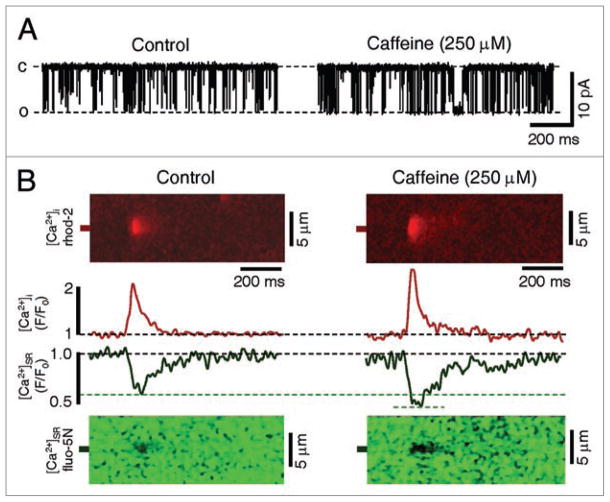
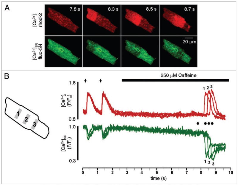
Cardiac contraction during systole is dependent on action potential-triggered Ca(2+) release from the sarcoplasmic reticulum (SR) through ryanodine receptor (RyR) channels. SR Ca(2+) release can also occur spontaneously during diastole, which causes a decrease in Ca(2+) content within the SR and contributes to arrhythmogenesis. Here, we use measurements of cytosolic Ca(2+) and intra-SR Ca(2+) ([Ca(2+)](SR)) to examine how RyR sensitization alters spontaneous SR Ca(2+) release events in rabbit ventricular myocytes. RyR sensitization with caffeine (250 microM) increased the open probability of single RyR channels, increased the initial frequency and amplitude of local SR Ca(2+) release events (Ca(2+) sparks), and decreased the [Ca(2+)](SR) level where Ca(2+) sparks terminated. In intact myocytes, caffeine applied during rest after steady-state electrical stimulation increased the frequency of spontaneous Ca(2+) waves and decreased the [Ca(2+)](SR) level where waves terminated. These effects caused a marked loss of SR Ca(2+) content. Therefore, increasing RyR activity has complex effects on cardiac function. Increased RyR activity during systole is beneficial as it increases SR Ca(2+) release and contractile strength. However, increased RyR activity during diastole produces spontaneous, arrhythmogenic Ca(2+) release events that lower SR Ca(2+) content and subsequently decrease contractility.
Figures


Similar articles
-
Alteration of sarcoplasmic reticulum Ca2+ release termination by ryanodine receptor sensitization and in heart failure.J Physiol. 2009 Nov 1;587(Pt 21):5197-209. doi: 10.1113/jphysiol.2009.177576. Epub 2009 Sep 7. J Physiol. 2009. PMID: 19736296 Free PMC article.
-
A novel mechanism of tandem activation of ryanodine receptors by cytosolic and SR luminal Ca2+ during excitation-contraction coupling in atrial myocytes.J Physiol. 2017 Jun 15;595(12):3835-3845. doi: 10.1113/JP273611. Epub 2017 Feb 1. J Physiol. 2017. PMID: 28028837 Free PMC article.
-
Ryanodine receptor current amplitude controls Ca2+ sparks in cardiac muscle.Circ Res. 2012 Jun 22;111(1):28-36. doi: 10.1161/CIRCRESAHA.112.265652. Epub 2012 May 24. Circ Res. 2012. PMID: 22628577 Free PMC article.
-
From the ryanodine receptor to cardiac arrhythmias.Circ J. 2009 Sep;73(9):1561-7. doi: 10.1253/circj.cj-09-0478. Epub 2009 Aug 10. Circ J. 2009. PMID: 19667488 Review.
-
Impact of RyR2 potentiation on myocardial function.Am J Physiol Heart Circ Physiol. 2017 Jun 1;312(6):H1105-H1109. doi: 10.1152/ajpheart.00855.2016. Epub 2017 Apr 7. Am J Physiol Heart Circ Physiol. 2017. PMID: 28389603 Review.
Cited by
-
Obstruction of ventricular Ca2+ -dependent arrhythmogenicity by inositol 1,4,5-trisphosphate-triggered sarcoplasmic reticulum Ca2+ release.J Physiol. 2018 Sep;596(18):4323-4340. doi: 10.1113/JP276319. Epub 2018 Aug 7. J Physiol. 2018. PMID: 30004117 Free PMC article.
-
Intercellular Ca(2+) waves: mechanisms and function.Physiol Rev. 2012 Jul;92(3):1359-92. doi: 10.1152/physrev.00029.2011. Physiol Rev. 2012. PMID: 22811430 Free PMC article. Review.
-
Elementary calcium signaling in arterial smooth muscle.Channels (Austin). 2019 Dec;13(1):505-519. doi: 10.1080/19336950.2019.1688910. Channels (Austin). 2019. PMID: 31797713 Free PMC article. Review.
-
β-Adrenergic stimulation increases the intra-sarcoplasmic reticulum Ca2+ threshold for Ca2+ wave generation.J Physiol. 2012 Dec 1;590(23):6093-108. doi: 10.1113/jphysiol.2012.236117. Epub 2012 Sep 17. J Physiol. 2012. PMID: 22988136 Free PMC article.
-
Dantrolene prevents arrhythmogenic Ca2+ release in heart failure.Am J Physiol Heart Circ Physiol. 2012 Feb 15;302(4):H953-63. doi: 10.1152/ajpheart.00936.2011. Epub 2011 Dec 16. Am J Physiol Heart Circ Physiol. 2012. PMID: 22180651 Free PMC article.
References
Publication types
MeSH terms
Substances
Grants and funding
LinkOut - more resources
Full Text Sources
Medical
Research Materials
Miscellaneous