. 2010 Feb;31(2):175-83.
doi: 10.1038/aps.2009.190.

Protective role of PI3-kinase/Akt/eNOS signaling in mechanical stress through inhibition of p38 mitogen-activated protein kinase in mouse lung

Affiliations

Protective role of PI3-kinase/Akt/eNOS signaling in mechanical stress through inhibition of p38 mitogen-activated protein kinase in mouse lung

Xin-qi Peng et al. Acta Pharmacol Sin. 2010 Feb.

Abstract

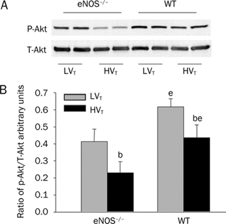
Aim: To test the hypothesis that PI3K/Akt/eNOS signaling has a protective role in a murine model of ventilation associated lung injury (VALI) through down-regulation of p38 MAPK signaling.

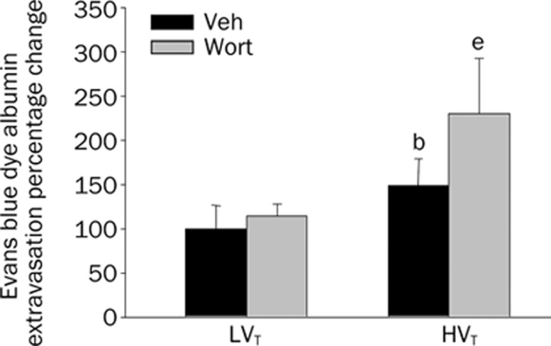
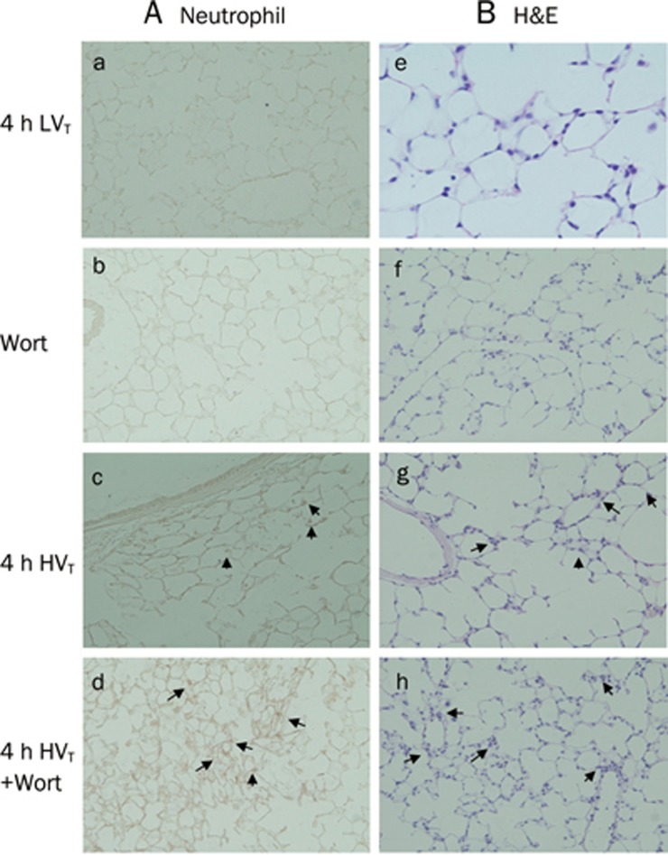
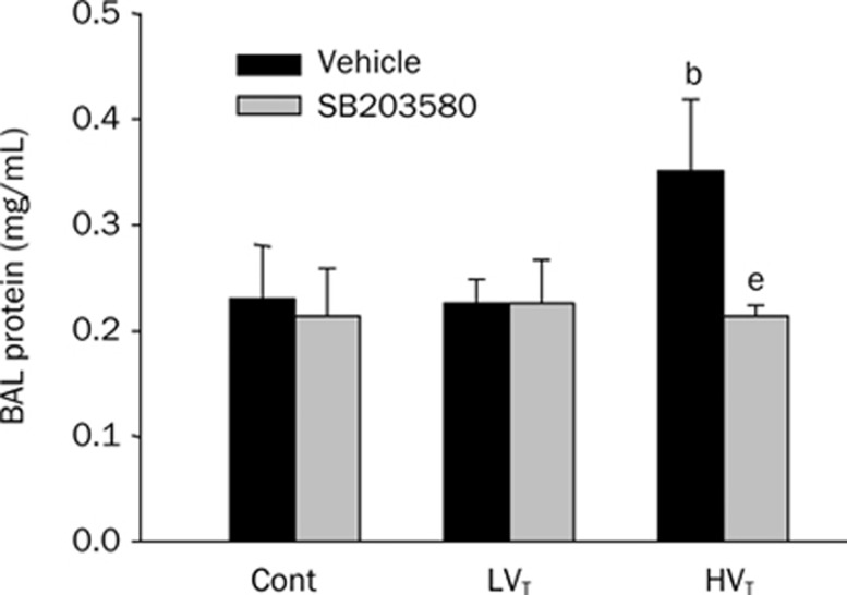
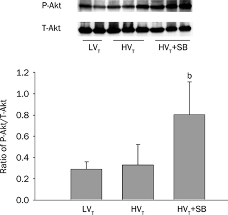
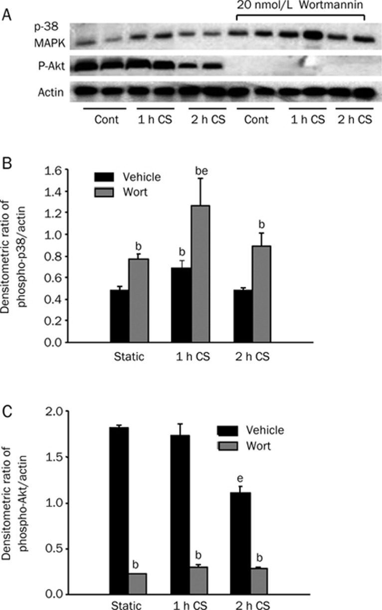
Methods: Male C57BL/J6 (wild-type, WT) or eNOS knockout mice (eNOS(-/-)) were exposed to mechanical ventilation (MV) with low (LV(T), 7 mL/kg) and high tidal volume (HV(T), 20 mL/kg) for 0-4 h. A subset of WT mice was administered the specific inhibitors of PI3K (100 nmol/L Wortmannin [Wort], ip) or of p38 MAPK (SB203580, 2 mg/kg, ip) 1 h before MV. Cultured type II alveolar epithelial cells C10 were exposed to 18% cyclic stretch for 2 h with or without 20 nmol/L Wort pretreatment. At the end of the experiment, the capillary leakage in vivo was assessed by extravasation of Evans blue dye (EBD), wet/dry weight ratio and lung lavage protein concentration. The lung tissue and cell lysate were also collected for protein and histological review.

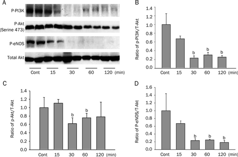
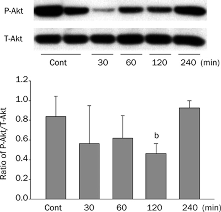
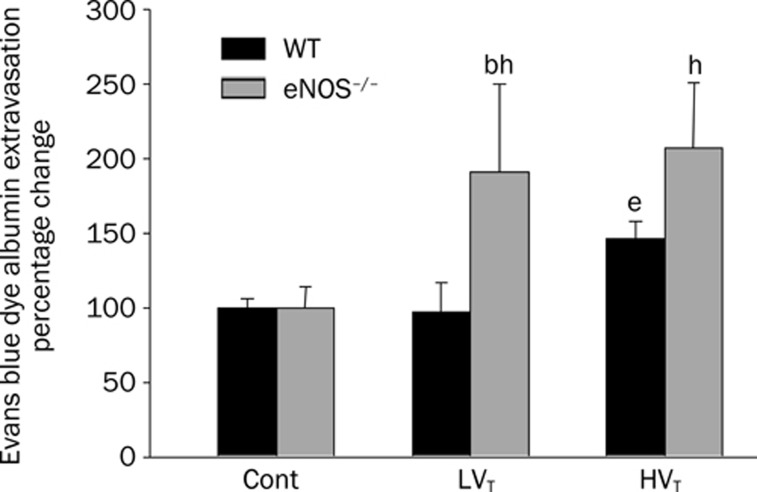
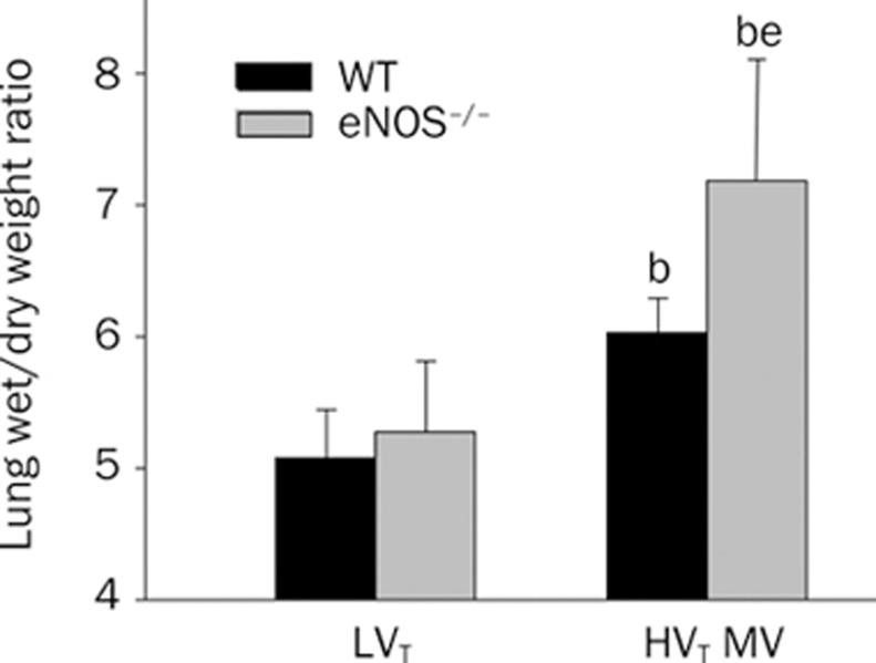
Results: MV decreased PI3K/Akt phosphorylation and eNOS expression but increased phospho-p38 MAPK expression along with a lung leakage of EBD. Inhibitions of phospho-Akt by Wort worsen the lung edema, whereas inhibition of p38 MAPK kinase restored activation of Akt together with alleviated capillary leakage. eNOS(-/-) mice showed an exacerbated lung edema and injury. The stretched C10 cells demonstrated that Wort diminished the activation of Akt, but potentiated phosphorylation of MAPK p38.

Conclusion: Our results indicate that PI-3K/Akt/eNOS pathway has significant protective effects in VALI by preventing capillary leakage, and that there is a cross-talk between PI3K/Akt and p38 MAPK pathways in vascular barrier dysfunction resulting from VALI.


Figures

Figure 1
Figure 1
The signaling pathway of PI3K/Akt/eNOS in response to MV. C57BL/6J mice were randomly exposed to spontaneous breathing (Control, Cont) or HVT (20 mL/kg) for 2 h. (A) Immunoblotting of WT mouse lung homogenates shows a decrease in PI3K, Akt, and eNOS phosphorylation after initiation of HVT MV detected by their active forms of antibody. (B), (C), and (D) show the densitometry of phospho-PI3K, phopho-AKT, and phospho-eNOS normalized to total Akt. n=3–4 mice per condition, bP<0.05 vs control. Note: the Figure 1A P-Akt bands represented a combination of the results came from several experiments.
Figure 2
Figure 2
Down-regulation of phospho-Akt in response to mechanical stress in a time-dependent fashion. (A) In a separate experiment of 4 h HVT MV, we observed that phospho-Akt at its serine residue 473 decreased most noticeably at 30 min and then gradually returned to control level at 2 h. (B) shows the densitometry of phopho-Akt normalized to total Akt. n=3–4 mice per condition, bP <0.05.
Figure 3
Figure 3
eNOS defective mice are less tolerant than WT mice in response to high amplitude mechanical stress featured with severe lung edema. Pulmonary vascular leakage was assessed at 4 h by the extravasation of evans blue dye(EBD) albumin in the lung parenchyma. MV with LVT caused no change in EBD influx in C57BL/6J mouse lungs, but caused a marked increase of EBD extravasation in eNOS−/− mouse lungs. In addition, HVT MV caused a 100% increase of EBD in eNOS−/− mouse lung in comparison with a 50% increase of EBA in WT mice. n=4 mice/experimental condition. bP<0.05 vs Control; eP<0.05 vs LVT; hP<0.05 vs WT vent-exposed mice.
Figure 4
Figure 4
Effects of eNOS deficiency on pulmonary vascular leakage during mechanical ventilation. As a separate measure of pulmonary edema, wet/dry lung weight ratios were determined at the end of 4 h HVT MV. HVT MV caused an increase of the lung wet-dry weight ratio especially in eNOS-deficient mice. n=4 mice/experimental condition. bP<0.05 vs LVT exposed animals; eP<0.05 compared with WT mice exposed to HVT MV.
Figure 5
Figure 5
Lower levels of phosph-Akt expression in eNOS−/− mice compared with WT mice after exposure to mechanical stress. We specifically evaluated the level of phosphor-Akt, a survivor signal in response to a variety of stresses, in our 4-h VALI model by immunoblotting. (A) Consistent with our data above, decreased phopho-Akt was blunted in WT mice at the end of 4 h HVT MV, while eNOS−/− mice showed a significant further decrease of phosphorylated Akt in response to 4 h HVT over the attenuated expression of phospho-Akt when exposed to LVT. (B) Densitometry of phopho-Akt normalized to total Akt. n=3–4 mice per condition. bP<0.05 vs LVT; eP<0.05 vs eNOS−/− mice.
Figure 6
Figure 6
Wortmannin (Wort) pretreatment aggravates the pulmonary vascular leakage in WT mice induced by 4 h HVT MV. C57BL/6J mice (WT) were pretreated with Wort (100 nmol/L, given intraperitoneally), a selective PI3Kase inhibitor, 1 h prior to exposure to MV. Wort itself dose not have effect on EBD assay (data not shown). Exposure of animals to LVT on the other hand caused no changes in these values as compared to controls (data not shown). However, Wort pretreatment significantly enhanced EBD lung accumulation in C57BL/6J mice caused by 4 h HVT MV. n=4–6 mice/experimental condition. bP<0.05 vs LVT; eP<0.05 vs HVT MV.
Figure 7
Figure 7
The morphological review of the lungs after pretreatment with Wort and mechanical ventilation. (A) Neutrophil immunostaining: the presence of parenchymal neutrophil was assessed using specific anti-neutrophil antibody. Exposure to 4 h HVT MV produced an increased staining of neutrophils in the interstitium and alveolus in WT mice (c, arrow) compared to LVT and Wort only (a and b). However, Wort — a specific inhibitor of PI3K--pretreatment exacerbated this effect (d, arrow). (B) Hematoxyline and eosin stain of lung tissue (×40): the presence of neutrophil infiltration in WT mice lungs when exposed to HVT for 4 h is similar as seen on the neutrophil immuno-staining but with occasional alveolar hemorrhage (g, arrow) compared to LVT and Wort only (e and f); Wort worsened the lung injury compared to that in animals ventilated alone (h, arrow). There were no striking histological changes in lungs of C57BL/6J mice exposed to LVT as compared with control animals (results not shown).
Figure 8
Figure 8
Inhibition of p38 MAP kinase prevents protein leakage in response to HVT MV. Mice were treated with the p38 MAP kinase inhibitor SB203580 (2 mg/kg, ip) 1 h prior to exposure to 1–2 h HVT MV. The data showed that the lung capillary leakage was attenuated by using the index of BAL protein at the end of 2 h HVT MV. n=4. bP<0.05 vs LVT; eP<0.05 vs 2 h HVT MV.
Figure 9
Figure 9
The effect of SB203580(SB) on the activation of Akt in WT mice exposed to 4 h mechanical ventilation. Western blotting on the mice lung exposed to HVT MV for 4 h demonstrated that the inhibition of p38 MAPK with SB203580 increased the activation of Akt (serine 473 phosphorylation), indicating interplay between these two signal pathways. n=3–4 mice per condition. bP<0.05.
Figure 10
Figure 10
The effect of PI3K inhibitor, 20 nmol/L Wort, on the expression of phospho-Akt and phospho-p38 MAPK. Pharmacologic antagonism of PI3K with Wort diminished the activation of Akt, but potentiated phosphorylation of MAPK p38 in C10, type II alveolar epithelial cells when exposed to 18% cyclic stretch (CS) from 1–2 h. A) the representative blotting; B and C) the densitometric ratio of phospho-p38/actin and phospho-Akt/actin. n=3–4. bP<0.05 vs Vehicle; eP<0.05 vs static. Note: the P-Akt bands represented a combination of the results came from several experiments.

References

    1. Ware LB, Matthay MA. The acute respiratory distress syndrome. N Engl J Med. 2000;342:1334–49. - PubMed
    1. Peng X, Hassoun PM, Sammani S, McVerry BJ, Burne MJ, Rabb H, et al. Protective effects of sphingosine 1-phosphate in murine endotoxin-induced inflammatory lung injury. Am J Respir Crit Care Med. 2004;169:1245–51. - PubMed
    1. Miyahara T, Hamanaka K, Weber DS, Drake DA, Anghelescu M, Parker JC. Phosphoinositide 3-kinase, Src, and Akt modulate acute ventilation-induced vascular permeability increases in mouse lungs. Am J Physiol Lung Cell Mol Physiol. 2007;293:L11–21. - PubMed
    1. Dimmeler S, Fleming I, Fisslthaler B, Hermann C, Busse R, Zeiher AM. Activation of nitric oxide synthase in endothelial cells by Akt-dependent phosphorylation. Nature. 1999;399:601–5. - PubMed
    1. Go YM, Boo YC, Park H, Maland MC, Patel R, Pritchard KA Jr, et al. Protein kinase B/Akt activates c-Jun NH2-terminal kinase by increasing NO production in response to shear stress. J Appl Physiol. 2001;91:1574–81. - PubMed

Publication types

Substances