. 2010 Feb 25;463(7284):1096-100.
doi: 10.1038/nature08735. Epub 2010 Feb 7.

Tbx3 improves the germ-line competency of induced pluripotent stem cells

Affiliations

Tbx3 improves the germ-line competency of induced pluripotent stem cells

Jianyong Han et al. Nature. .

Abstract

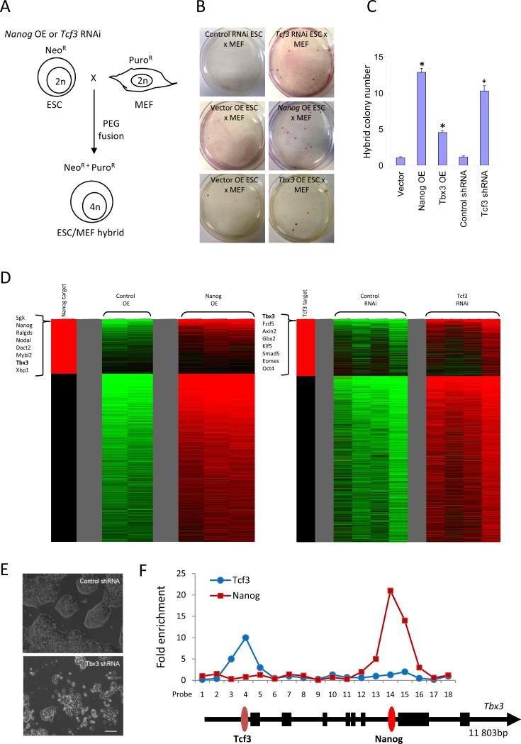
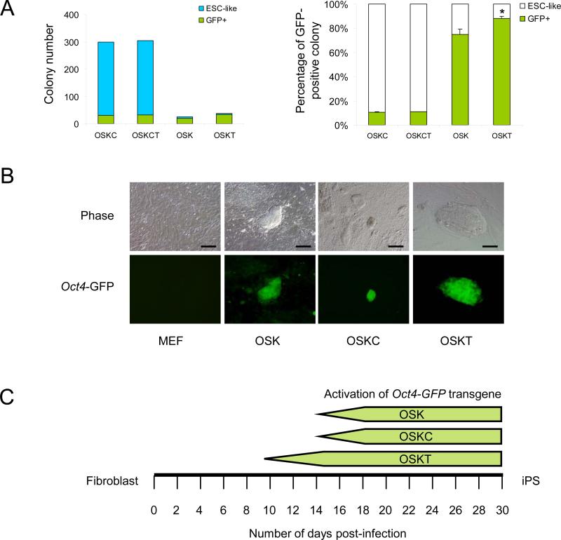
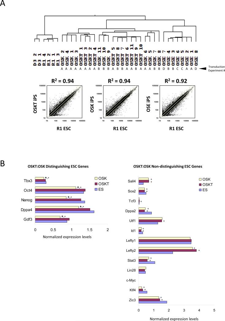
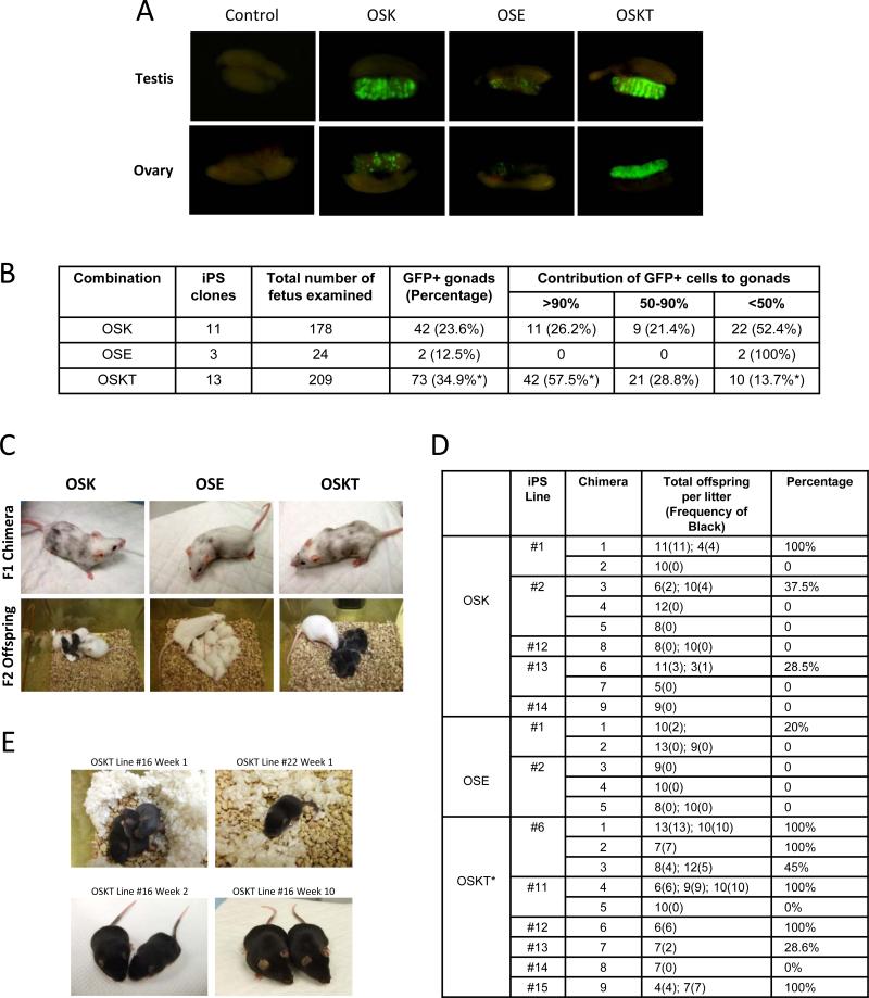
Induced pluripotent stem (iPS) cells can be obtained by the introduction of defined factors into somatic cells. The combination of Oct4 (also known as Pou5f1), Sox2 and Klf4 (which we term OSK) constitutes the minimal requirement for generating iPS cells from mouse embryonic fibroblasts. These cells are thought to resemble embryonic stem cells (ESCs) on the basis of global gene expression analyses; however, few studies have tested the ability and efficiency of iPS cells to contribute to chimaerism, colonization of germ tissues, and most importantly, germ-line transmission and live birth from iPS cells produced by tetraploid complementation. Using genomic analyses of ESC genes that have roles in pluripotency and fusion-mediated somatic cell reprogramming, here we show that the transcription factor Tbx3 significantly improves the quality of iPS cells. iPS cells generated with OSK and Tbx3 (OSKT) are superior in both germ-cell contribution to the gonads and germ-line transmission frequency. However, global gene expression profiling could not distinguish between OSK and OSKT iPS cells. Genome-wide chromatin immunoprecipitation sequencing analysis of Tbx3-binding sites in ESCs suggests that Tbx3 regulates pluripotency-associated and reprogramming factors, in addition to sharing many common downstream regulatory targets with Oct4, Sox2, Nanog and Smad1. This study underscores the intrinsic qualitative differences between iPS cells generated by different methods, and highlights the need to rigorously characterize iPS cells beyond in vitro studies.


Figures

Figure 1
Figure 1
Global gene expression profiling reveals Tbx3 aids cell fusion-mediated reprogramming. (A) Modified ESCs with Nanog over-expression (OE) or Tcf3 RNAi were fused with MEFs to generate tetraploid ESC/MEF hybrids resistant to neomycin and puromycin. (B) Nanog OE, Tbx3 OE and Tcf3 RNAi enhanced cell fusion-mediated reprogramming of MEFs. Representative examples illustrate the emergence of ESC/MEF hybrid colonies. Control ESC fusion with MEFs resulted in an average of one per experiment whereas Tcf3 RNAi, Nanog OE or Tbx3 OE ESCs produced numerous hybrid clones. (C) Nanog OE ESCs were efficient in reprogramming MEFs, generating 13 colonies, followed by Tcf3 RNAi (10) and Tbx3 OE (4.5). The numbers represent the average of four independent fusion experiments. * denotes significantly different from vector, + denotes significantly different from control shRNA; error bars represent s.e.m. (D) The heat-map shows all genes which were increased in treated ESCs compared to controls. Tbx3 was among the most highly up-regulated genes in Nanog OE and Tcf3 RNAi ESCs. The left-most column in red indicates direct gene targets of Nanog or Tcf3 based on the ChIP-PET and ChIP-chip databases respectively. (E) RNAi knockdown of Tbx3 in ESCs led to a loss of self-renewal and induced differentiation. Scale bar = 100 μm. (F) Enrichment of Tcf3 and Nanog occupancy on the Tbx3 gene, as measured by ChIP-qPCR.
Figure 2
Figure 2
Generation of iPS cells with Oct4, Sox2, Klf4 and Tbx3 retroviral transduction. (A) OSKC and OSKCT induced ~300 ESC-like colonies with ~10% expressing GFP, whereas OSKT induced an average of 38 compared to 26 ESC-like colonies from OSK, with 89% and 74% GFP activation respectively. Colonies were assessed and counted on day 16. Data represents the average of three independent transduction experiments. * denotes significantly different from OSK; p<0.05; error bars represent s.e.m. (B) OSK and OSKT iPS cell clones showed tight, domed-shaped ESC-like morphology and uniform GFP expression throughout the colony, whereas OSKC clones appeared as flattened, transformed cells with sparse GFP expression. Scale bar = 100 μm. (C) OSKT induced the activation of Oct4-GFP transgene in iPS derived from primary MEFs at 9-10 days post-infection while OSK and OSKC combinations required 14 days observed in four independent transduction experiment.
Figure 3
Figure 3
Transcriptome of iPS cell clones generated with different combinations of reprogramming factors. (A) Hierarchical clustering of global expression profiling showed that OSKT and OSK clones are more similar to wild-type ESCs than OSE, but indistinguishable from each other based on correlation coefficient. R2 value was obtained from the average individual gene signal intensity of all iPS clones in each combination and compared against R1 ESCs. The independent transduction experiments where the clones were isolated are denoted as A, B, C, and D. (B) Analysis of individual ESC-associated gene profiles revealed a subset that could distinguish OSKT from OSK-derived iPS cells. ‘Distinguishing’ ESC genes were expressed at levels similar between OSKT and ESCs but significantly lower in OSK. The majority of other ESC-associated genes were ‘non-distinguishing’ and present at levels similar between both OSKT and OSK. * denotes significantly different from OSKT; + denotes significantly different from ESC; p<0.05. Changes in gene expression based on microarray were confirmed with qPCR (Figure S11).
Figure 4
Figure 4
OSKT iPS cells show enhanced germ-line contribution and transmission. The iPS clone ID and the types of analyses that were performed is summarized in Table S7. (A) Representative photos showing the quantitative contribution and spatial distribution of GFP+ cells generated with different reprogramming factor combinations in the gonads of chimeric embryos. (B) Tabulation comparing the contribution of GFP+ iPS cell-derived germ cells to the gonads of chimeric fetuses and their spatial distribution with different combinations of factors. OSKT iPS cells were most effective at colonizing the gonads, compared to OSK and OSE iPS cells. * denotes significantly different from OSK, p<0.01. At least eleven independent sets of microinjections were performed for OSK versus OSKT comparison. (C) Representative photos showing the contribution of iPS cells to chimeric coat and the production of F2 offspring after crossing with albino mice. Black offspring indicates germ-line transmission of iPS cells. (D) Table summarizing the germ-line transmission frequency for iPS cells generated with the different combination of factors. For each combination, between two to six iPS cell lines were used to obtain live chimeras, of which, at least one male from each combination were crossed with an albino female to determine the frequency of black F2. * denotes significantly different from OSK and OSE, p<0.005 and p<0.001 respectively. (E) Live birth animals obtained from two OSKT iPS cell lines tested by tetraploid complementation.

Similar articles

Cited by

References

    1. Takahashi K, Yamanaka S. Induction of pluripotent stem cells from mouse embryonic and adult fibroblast cultures by defined factors. Cell. 2006;126:663–76. - PubMed
    1. Huangfu D, et al. Induction of pluripotent stem cells by defined factors is greatly improved by small-molecule compounds. Nat Biotechnol. 2008;26:795–7. - PMC - PubMed
    1. Shi Y, et al. Induction of pluripotent stem cells from mouse embryonic fibroblasts by Oct4 and Klf4 with small-molecule compounds. Cell Stem Cell. 2008;3:568–74. - PubMed
    1. Silva J, et al. Promotion of reprogramming to ground state pluripotency by signal inhibition. PLoS Biol. 2008;6:e253. - PMC - PubMed
    1. Okita K, Nakagawa M, Hyenjong H, Ichisaka T, Yamanaka S. Generation of mouse induced pluripotent stem cells without viral vectors. Science. 2008;322:949–53. - PubMed

Publication types

MeSH terms

Associated data