

Increased expression of human T-cell immunoglobulin- and mucin-domain-containing molecule-4 in peripheral blood mononuclear cells from patients with system lupus erythematosus
- PMID: 20140011
- PMCID: PMC4001890
- DOI: 10.1038/cmi.2009.118
Increased expression of human T-cell immunoglobulin- and mucin-domain-containing molecule-4 in peripheral blood mononuclear cells from patients with system lupus erythematosus
Abstract
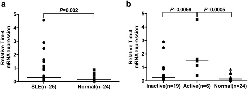
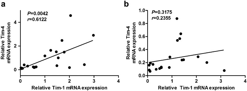
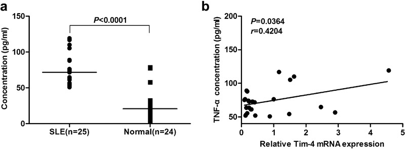
Systemic lupus erythematosus (SLE) is a prototypic autoimmune disease. Innate and adaptive immunity cooperatively contribute to the development of SLE. Antigen-presenting cells (APCs) have been suggested to link innate and adaptive immunity. T-cell immunoglobulin- and mucin-domain-containing molecule-4 (Tim-4; also known as Timd4), expressed primarily on the surface of APCs, is a member of the TIM family, a recently described group of molecules that have received much attention as potential regulators of the immune system. In this study, we used quantitative real-time reverse transcription-polymerase chain reaction to examine the mRNA expression of Tim-4 in peripheral blood mononuclear cells (PBMCs) from SLE patients and further analyzed the correlation between the expression of Tim-4 and Tim-1 (a potential ligand for Tim-4) in PBMCs and serum tumor necrosis factor (TNF)-alpha levels. The results showed that Tim-4 mRNA expression in PBMCs was significantly higher in SLE patients than in healthy controls, especially those patients in the active phase of disease. Moreover, Tim-4 mRNA levels were closely correlated with Tim-1 mRNA levels in PBMCs and with serum TNF-alpha levels in SLE patients but not in the control group. Taken together, these results demonstrate that Tim-4 may be involved in the pathogenesis of SLE.
Figures
Similar articles
-
Increased Tim-3 expression on peripheral T lymphocyte subsets and association with higher disease activity in systemic lupus erythematosus.Diagn Pathol. 2015 Jun 16;10:71. doi: 10.1186/s13000-015-0306-0. Diagn Pathol. 2015. PMID: 26076826 Free PMC article.
-
Expression of human T cell immunoglobulin domain and mucin-3 (TIM-3) and TIM-3 ligands in peripheral blood from patients with systemic lupus erythematosus.Arch Dermatol Res. 2016 Oct;308(8):553-61. doi: 10.1007/s00403-016-1665-4. Epub 2016 Jul 9. Arch Dermatol Res. 2016. PMID: 27394439
-
Expression of human TIM-1 and TIM-3 on lymphocytes from systemic lupus erythematosus patients.Scand J Immunol. 2008 Jan;67(1):63-70. doi: 10.1111/j.1365-3083.2007.02038.x. Scand J Immunol. 2008. PMID: 18052965
-
TIM-3 as a new therapeutic target in systemic lupus erythematosus.Mol Biol Rep. 2010 Jan;37(1):395-8. doi: 10.1007/s11033-009-9833-7. Mol Biol Rep. 2010. PMID: 19768575 Review.
-
The T-cell immunoglobulin and mucin domain (Tim) gene family in asthma, allergy, and autoimmunity.Allergy Asthma Proc. 2013 Jan-Feb;34(1):e21-6. doi: 10.2500/aap.2013.34.3646. Allergy Asthma Proc. 2013. PMID: 23406933 Review.
Cited by
-
Tim-4 protects mice against lipopolysaccharide-induced endotoxic shock by suppressing the NF-κB signaling pathway.Lab Invest. 2016 Nov;96(11):1189-1197. doi: 10.1038/labinvest.2016.94. Epub 2016 Sep 12. Lab Invest. 2016. PMID: 27617399
-
Function and characteristics of TIM‑4 in immune regulation and disease (Review).Int J Mol Med. 2023 Feb;51(2):10. doi: 10.3892/ijmm.2022.5213. Epub 2022 Dec 16. Int J Mol Med. 2023. PMID: 36524355 Free PMC article. Review.
-
IRG1/itaconate enhances efferocytosis by activating Nrf2-TIM4 signaling pathway to alleviate con A induced autoimmune liver injury.Cell Commun Signal. 2025 Feb 5;23(1):63. doi: 10.1186/s12964-025-02075-5. Cell Commun Signal. 2025. PMID: 39910615 Free PMC article.
-
TIM-4 promotes the growth of non-small-cell lung cancer in a RGD motif-dependent manner.Br J Cancer. 2015 Nov 17;113(10):1484-92. doi: 10.1038/bjc.2015.323. Epub 2015 Oct 29. Br J Cancer. 2015. PMID: 26512878 Free PMC article.
-
Significance of K(L/V)WX(I/L/V)P Epitope of the B2Gpi in Its (Patho)Physiologic Function.EJIFCC. 2011 Dec 22;22(4):118-24. eCollection 2011 Dec. EJIFCC. 2011. PMID: 27683401 Free PMC article.
References
-
- Zuñiga R, Ng S, Peterson MG, Reveille JD, Baethge BA, Alarcón GS, et al. Low-binding alleles of Fcgama receptor types IIA and IIIA are inherited independently and are associated with systemic lupus erythematosus in Hispanic patients. Arthritis Rheum. 2001;44:361–367. - PubMed
-
- Han JW, Zheng HF, Cui Y, Sun LD, Ye DQ, Hu Z, et al. Genome-wide association study in a Chinese Han population identifies nine new susceptibility loci for systemic lupus erythematosus. Nat Genet. 2009;41:1234–1237. - PubMed
-
- Xiang YJ, Dai SM. Prevalence of rheumatic diseases and disability in China. Rheumatol Int. 2009;29:481–490. - PubMed
-
- Agarwal S, Elliott JR, Manzi S. Atherosclerosis risk factors in systemic lupus erythematosus. Curr Rheumatol Rep. 2009;11:241–247. - PubMed
Publication types
MeSH terms
Substances
LinkOut - more resources
Full Text Sources
Medical
Miscellaneous
