

Keratin promoter based gene manipulation in the murine conducting airway
- PMID: 20140084
- PMCID: PMC2815352
- DOI: 10.7150/ijbs.6.68
Keratin promoter based gene manipulation in the murine conducting airway
Abstract
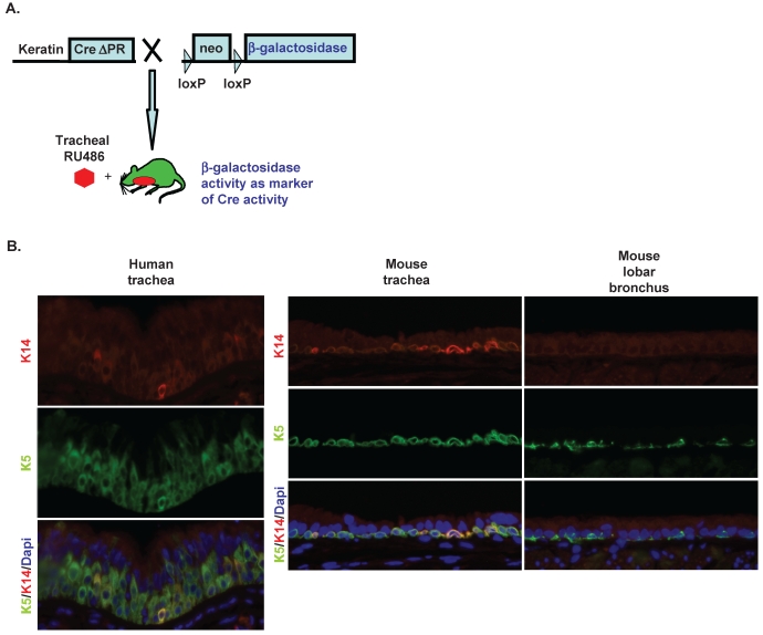
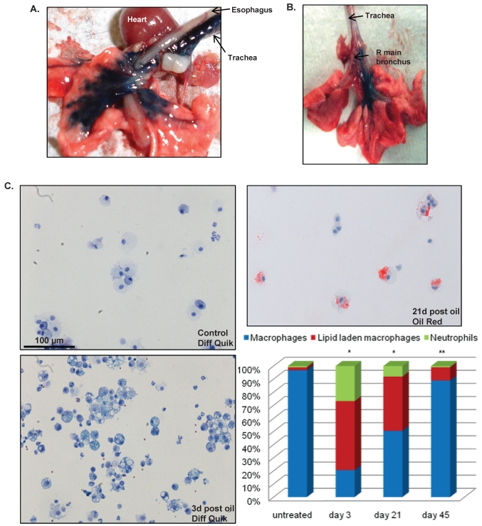
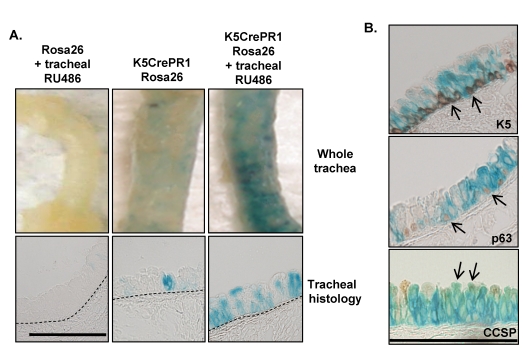
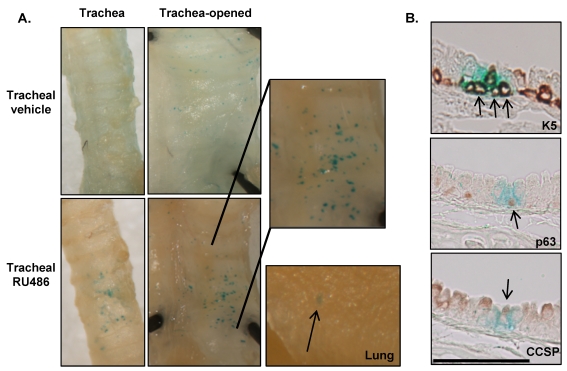
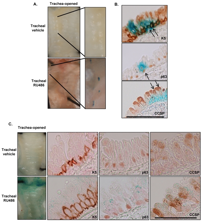
Systems capable of targeting genetic manipulations to keratin-positive airway basal cells are more poorly developed than systems targeting other airway epithelial cell populations and this has likely hindered development of animal models of diseases such as lung squamous cell carcinoma. Although keratin promoter driven-Cre recombinase constructs are potentially useful for targeting these cells, these constructs have substantially higher activity in the skin and oral epithelium than in the airways. We developed a method for delivering RU486, the conditional activator of Cre recombinase progesterone receptor (CrePR) fusion proteins to the lung and then examined the activity of three keratin-driven CrePR constructs in the conducting airways. We also developed a technique for survival bronchioalveolar lavage on non-ventilated animals to examine the effects of the acetone/oil vehicle required to deliver RU486 to the lung. K5CrePR1 and K14CrePR1 constructs differ only in the keratin promoter used to target CrePR1 expression while K5Cre*PR contains a truncated progesterone receptor designed to reduce RU486-independent Cre activity. While all three constructs demonstrate RU486-inducible Cre activity in the conducting airways, both construct activity and tightness of regulation vary considerably. K5Cre*PR is the most tightly regulated Cre driver making it ideal for targeting somatic mutations to the airway epithelia while K5CrePR1 and K14CrePR1 may be better suited to studying diseases of the conducting airways where gene targeting of keratin expressing cells and their derivatives is desired.
Keywords: Cre recombinase; Keratins; basal cells.
Conflict of interest statement
Conflicts of interest: The authors report no conflict of interest. The authors alone are responsible for the content and writing of this manuscript.
Figures
Similar articles
-
Inducible gene targeting in postnatal myocardium by cardiac-specific expression of a hormone-activated Cre fusion protein.Circ Res. 2001 Mar 30;88(6):587-92. doi: 10.1161/01.res.88.6.587. Circ Res. 2001. PMID: 11282892
-
RU486-inducible recombination in the salivary glands of lactoferrin promoter-driven green fluorescent Cre transgenic mice.Genesis. 2010 Oct 1;48(10):585-95. doi: 10.1002/dvg.20666. Genesis. 2010. PMID: 20715174
-
Neuron-specific and inducible recombination by Cre recombinase in the mouse.Neuroreport. 2008 Apr 16;19(6):621-4. doi: 10.1097/WNR.0b013e3282fb7d99. Neuroreport. 2008. PMID: 18382274
-
Overlapping but distinct profiles of gene expression elicited by glucocorticoids and progestins.Recent Prog Horm Res. 2003;58:199-226. doi: 10.1210/rp.58.1.199. Recent Prog Horm Res. 2003. PMID: 12795420 Review.
-
Conditional gene targeting on the pure C57BL/6 genetic background.Neurosci Res. 2007 Jun;58(2):105-12. doi: 10.1016/j.neures.2007.01.004. Epub 2007 Jan 18. Neurosci Res. 2007. PMID: 17298852 Review.
Cited by
-
Smad4 loss promotes lung cancer formation but increases sensitivity to DNA topoisomerase inhibitors.Oncogene. 2016 Feb 4;35(5):577-586. doi: 10.1038/onc.2015.112. Epub 2015 Apr 20. Oncogene. 2016. PMID: 25893305 Free PMC article.
-
Conditional knockout of ITGB4 in bronchial epithelial cells directs bronchopulmonary dysplasia.J Cell Mol Med. 2023 Dec;27(23):3760-3772. doi: 10.1111/jcmm.17948. Epub 2023 Sep 12. J Cell Mol Med. 2023. PMID: 37698050 Free PMC article.
-
Dynamic changes in intracellular ROS levels regulate airway basal stem cell homeostasis through Nrf2-dependent Notch signaling.Cell Stem Cell. 2014 Aug 7;15(2):199-214. doi: 10.1016/j.stem.2014.05.009. Epub 2014 Jun 19. Cell Stem Cell. 2014. PMID: 24953182 Free PMC article.
-
Role of PTEN in basal cell derived lung carcinogenesis.Mol Carcinog. 2014 Oct;53(10):841-6. doi: 10.1002/mc.22030. Epub 2013 Apr 26. Mol Carcinog. 2014. PMID: 23625632 Free PMC article.
-
Loss of transforming growth factor beta type II receptor increases aggressive tumor behavior and reduces survival in lung adenocarcinoma and squamous cell carcinoma.Clin Cancer Res. 2012 Apr 15;18(8):2173-83. doi: 10.1158/1078-0432.CCR-11-2557. Epub 2012 Mar 7. Clin Cancer Res. 2012. PMID: 22399565 Free PMC article.
References
-
- Kim CF, Jackson EL, Woolfenden AE. et al.Identification of bronchioalveolar stem cells in normal lung and lung cancer. Cell. 2005;121:823–35. - PubMed
Publication types
MeSH terms
Substances
Grants and funding
LinkOut - more resources
Full Text Sources
Molecular Biology Databases
Research Materials
