

Comprehensive identification and modified-site mapping of S-nitrosylated targets in prostate epithelial cells
- PMID: 20140087
- PMCID: PMC2816712
- DOI: 10.1371/journal.pone.0009075
Comprehensive identification and modified-site mapping of S-nitrosylated targets in prostate epithelial cells
Abstract
Background: Although overexpression of nitric oxide synthases (NOSs) has been found associated with prostate diseases, the underlying mechanisms for NOS-related prostatic diseases remain unclear. One proposed mechanism is related to the S-nitrosylation of key regulatory proteins in cell-signaling pathways due to elevated levels of NO in the prostate. Thus, our primary objective was to identify S-nitrosylated targets in an immortalized normal prostate epithelial cell line, NPrEC.
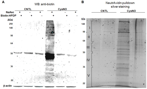
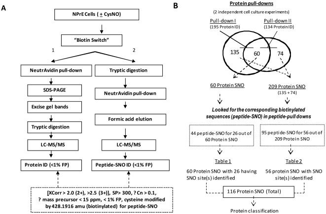
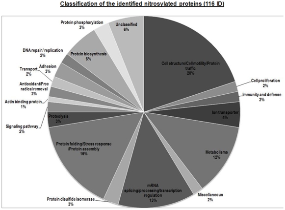
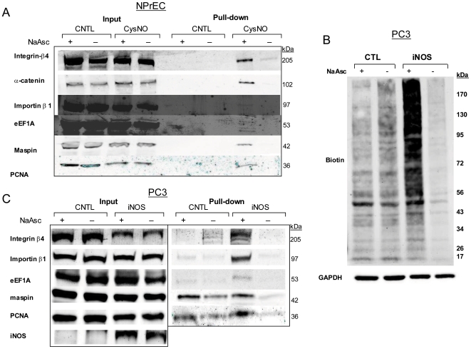
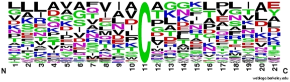
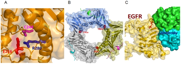
Methodology/principal findings: We treated NPrEC with nitroso-cysteine and used the biotin switch technique followed by gel-based separation and mass spectrometry protein identification (using the LTQ-Orbitrap) to discover S-nitrosylated (SNO) proteins in the treated cells. In parallel, we adapted a peptide pull-down methodology to locate the site(s) of S-nitrosylation on the protein SNO targets identified by the first technique. This combined approach identified 116 SNO proteins and determined the sites of modification for 82 of them. Over 60% of these proteins belong to four functional groups: cell structure/cell motility/protein trafficking, protein folding/protein response/protein assembly, mRNA splicing/processing/transcriptional regulation, and metabolism. Western blot analysis validated a subset of targets related to disease development (proliferating cell nuclear antigen, maspin, integrin beta4, alpha-catenin, karyopherin [importin] beta1, and elongation factor 1A1). We analyzed the SNO sequences for their primary and secondary structures, solvent accessibility, and three-dimensional structural context. We found that about 80% of the SNO sites that can be mapped into resolved structures are buried, of which approximately half have charged amino acids in their three-dimensional neighborhood, and the other half residing within primarily hydrophobic pockets.
Conclusions/significance: We here identified 116 potential SNO targets and mapped their putative SNO sites in NPrEC. Elucidation of how this post-translational modification alters the function of these proteins should shed light on the role of NO in prostate pathologies. To our knowledge, this is the first report identifying SNO targets in prostate epithelial cells.
Conflict of interest statement
Figures
Similar articles
-
Site-mapping of in vitro S-nitrosation in cardiac mitochondria: implications for cardioprotection.Mol Cell Proteomics. 2011 Mar;10(3):M110.004721. doi: 10.1074/mcp.M110.004721. Epub 2010 Oct 29. Mol Cell Proteomics. 2011. PMID: 21036925 Free PMC article.
-
Functional proteomics approaches for the identification of transnitrosylase and denitrosylase targets.Methods. 2013 Aug 1;62(2):151-60. doi: 10.1016/j.ymeth.2013.02.002. Epub 2013 Feb 18. Methods. 2013. PMID: 23428400 Free PMC article.
-
SNOSID, a proteomic method for identification of cysteine S-nitrosylation sites in complex protein mixtures.Proc Natl Acad Sci U S A. 2006 Jan 24;103(4):1012-7. doi: 10.1073/pnas.0508412103. Epub 2006 Jan 17. Proc Natl Acad Sci U S A. 2006. PMID: 16418269 Free PMC article.
-
Methodologies for the characterization, identification and quantification of S-nitrosylated proteins.Biochim Biophys Acta. 2012 Jun;1820(6):675-83. doi: 10.1016/j.bbagen.2011.03.013. Epub 2011 Apr 5. Biochim Biophys Acta. 2012. PMID: 21440604 Free PMC article. Review.
-
Screening systems for the identification of S-nitrosylated proteins.Nitric Oxide. 2011 Aug 1;25(2):108-11. doi: 10.1016/j.niox.2010.11.002. Epub 2010 Nov 24. Nitric Oxide. 2011. PMID: 21111056 Review.
Cited by
-
Nitric Oxide Mediated Transcriptome Profiling Reveals Activation of Multiple Regulatory Pathways in Arabidopsis thaliana.Front Plant Sci. 2016 Jun 29;7:975. doi: 10.3389/fpls.2016.00975. eCollection 2016. Front Plant Sci. 2016. PMID: 27446194 Free PMC article.
-
The S-nitrosylation status of PCNA localized in cytosol impacts the apoptotic pathway in a Parkinson's disease paradigm.PLoS One. 2015 Feb 12;10(2):e0117546. doi: 10.1371/journal.pone.0117546. eCollection 2015. PLoS One. 2015. PMID: 25675097 Free PMC article.
-
Cysteine 96 of Ntcp is responsible for NO-mediated inhibition of taurocholate uptake.Am J Physiol Gastrointest Liver Physiol. 2013 Oct 1;305(7):G513-9. doi: 10.1152/ajpgi.00089.2013. Epub 2013 Jul 25. Am J Physiol Gastrointest Liver Physiol. 2013. PMID: 23886862 Free PMC article.
-
The DUSP26 phosphatase activator adenylate kinase 2 regulates FADD phosphorylation and cell growth.Nat Commun. 2014;5:3351. doi: 10.1038/ncomms4351. Nat Commun. 2014. PMID: 24548998 Free PMC article.
-
Identification of new targets of S-nitrosylation in neural stem cells by thiol redox proteomics.Redox Biol. 2020 May;32:101457. doi: 10.1016/j.redox.2020.101457. Epub 2020 Feb 7. Redox Biol. 2020. PMID: 32088623 Free PMC article.
References
-
- Fibbi B, Penna G, Morelli A, Adorini L, Maggi M. Chronic inflammation in the pathogenesis of benign prostatic hyperplasia. Int J Androl. In press 2009 - PubMed
-
- Sciarra A, Mariotti G, Salciccia S, Gomez AA, Monti S, et al. Prostate growth inflammation. J Steroid Biochem Mol Biol. 2008;108:254–260. - PubMed
-
- De Marzo AM, Nakai Y, Nelson WG. Inflammation, atrophy, and prostate carcinogenesis. Urol Oncol. 2007;25:398–400. - PubMed
-
- Aaltoma SH, Lipponen PK, Kosma VM. Inducible nitric oxide synthase (iNOS) expression and its prognostic value in prostate cancer. Anticancer Res. 2001;21:3101–3106. - PubMed
-
- Baltaci S, Orhan D, Gogus C, Turkolmez K, Tulunay O, et al. Inducible nitric oxide synthase expression in benign prostatic hyperplasia, low- and high-grade prostatic intraepithelial neoplasia and prostatic carcinoma. BJU Int. 2001;88:100–103. - PubMed
Publication types
MeSH terms
Substances
Grants and funding
LinkOut - more resources
Full Text Sources
