. 2010 Feb 5;5(2):e9079.
doi: 10.1371/journal.pone.0009079.

How cholesterol constrains glycolipid conformation for optimal recognition of Alzheimer's beta amyloid peptide (Abeta1-40)

Affiliations

How cholesterol constrains glycolipid conformation for optimal recognition of Alzheimer's beta amyloid peptide (Abeta1-40)

Nouara Yahi et al. PLoS One. .

Abstract

Membrane lipids play a pivotal role in the pathogenesis of Alzheimer's disease, which is associated with conformational changes, oligomerization and/or aggregation of Alzheimer's beta-amyloid (Abeta) peptides. Yet conflicting data have been reported on the respective effect of cholesterol and glycosphingolipids (GSLs) on the supramolecular assembly of Abeta peptides. The aim of the present study was to unravel the molecular mechanisms by which cholesterol modulates the interaction between Abeta(1-40) and chemically defined GSLs (GalCer, LacCer, GM1, GM3). Using the Langmuir monolayer technique, we show that Abeta(1-40) selectively binds to GSLs containing a 2-OH group in the acyl chain of the ceramide backbone (HFA-GSLs). In contrast, Abeta(1-40) did not interact with GSLs containing a nonhydroxylated fatty acid (NFA-GSLs). Cholesterol inhibited the interaction of Abeta(1-40) with HFA-GSLs, through dilution of the GSL in the monolayer, but rendered the initially inactive NFA-GSLs competent for Abeta(1-40) binding. Both crystallographic data and molecular dynamics simulations suggested that the active conformation of HFA-GSL involves a H-bond network that restricts the orientation of the sugar group of GSLs in a parallel orientation with respect to the membrane. This particular conformation is stabilized by the 2-OH group of the GSL. Correspondingly, the interaction of Abeta(1-40) with HFA-GSLs is strongly inhibited by NaF, an efficient competitor of H-bond formation. For NFA-GSLs, this is the OH group of cholesterol that constrains the glycolipid to adopt the active L-shape conformation compatible with sugar-aromatic CH-pi stacking interactions involving residue Y10 of Abeta(1-40). We conclude that cholesterol can either inhibit or facilitate membrane-Abeta interactions through fine tuning of glycosphingolipid conformation. These data shed some light on the complex molecular interplay between cell surface GSLs, cholesterol and Abeta peptides, and on the influence of this molecular ballet on Abeta-membrane interactions.


Conflict of interest statement

Competing Interests: The authors have declared that no competing interests exist.

Figures

Figure 1
Figure 1. Effects of cholesterol on the recognition of GalCer-HFA and GalCer-NFA by Aβ1–40.
a–c. Kinetics of Aβ1–40 insertion into a monolayer of natural GalCer-HFA (a), natural GalCer-NFA (b) or GalCer-C12 (c) in either the absence (▪) or presence of cholesterol (□). The pure GalCer and mixed GalCer/cholesterol (1∶1, mol:mol) monolayers were prepared at an initial surface pressure of 13–15 mN.m−1 as indicated in Materials and Methods. The data show the evolution of the surface pressure following the injection of Aβ1–40 (1 µM) in the aqueous phase underneath the monolayer. Each experiment was performed in triplicate and one representative curve is shown (S.D. <10%). The chemical structures of GalCer-HFA (with a C18:0 2-OH fatty acid), GalCer-NFA (with a C18:0 fatty acid) and GalCer-C12 (with a C12:0 fatty acid) are shown in the lower right panel. The stereochemistry of the C2 atom is indicated by an asterisk.
Figure 2
Figure 2. Effects of cholesterol on the recognition of synthetic LacCer (LacCer-C8) by Aβ1–40.
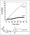
Upper panel: the kinetics of Aβ1–40 insertion into a monolayer of synthetic LacCer-C8 (with a C8:0 fatty acid) were measured in either the absence (▪) or presence of cholesterol (□). The pure LacCer and mixed LacCer/cholesterol (1∶1, mol:mol) monolayers were prepared at an initial surface pressure of 13–15 mN.m−1 as indicated in Materials and Methods. The data show the evolution of the surface pressure following the injection of Aβ1–40 (1 µM) in the aqueous phase underneath the monolayer. Each experiment was performed in triplicate and one representative curve is shown (S.D. <10%). The chemical structure of LacCer-C8 is shown in the lower panel.
Figure 3
Figure 3. Effects of cholesterol on the recognition of gangliosides GM3 and GM1 by Aβ1–40.
Kinetics of Aβ1–40 insertion into a monolayer of GM3 (a) or GM1(b) in either the absence (▪) or presence of cholesterol (□). The pure ganglioside and mixed ganglioside/cholesterol (1∶1, mol:mol) monolayers were prepared at an initial surface pressure of 13–15 mN.m−1 as indicated in Materials and Methods. The data show the evolution of the surface pressure following the injection of Aβ1–40 (1 µM) in the aqueous phase underneath the monolayer. Each experiment was performed in triplicate and one representative curve is shown (S.D. <10%). The chemical structures of GM3 and GM1 (with a C18:0 fatty acid) are shown in the lower panel. Note that the chemical structure of the glycone moiety of GM1 is derived from the one of GM3 (GM3, NeuAcα2-3Galβ1-4Glcβ1-Cer; GM1, Galα1-3GalNacβ1-4- (highlighted in red) branched on the second Gal residue of GM3. The common oligosaccharide part shared by GM3 and GM1 is highlighted in blue.
Figure 4
Figure 4. Molecular modeling simulations of GalCer-HFA and GalCer-NFA (alone or complexed with cholesterol).
Molecular dynamics simulations were performed as indicated in Materials and Methods. a- GalCer-NFA; b- GalCer-NFA complexed with cholesterol (in green); c- GalCer-HFA (the α-OH group is indicated); d- Superposition of GalCer-NFA (identical to a, colored green) with the GalCer-NFA/cholesterol complex (identical to b). Note the distinct orientation of the galactose headgroup (Gal); e- Superposition of GalCer-NFA (in green) with GalCer-HFA; f- In GalCer-HFA, the galactose head group is maintained in a shovel-like conformation by a network of H-bonds involving the NH of sphingosine (donor group) and the 2-OH of the fatty acid and the oxygen atom of the glycosidic bond (both acceptor groups); g- In GalCer-NFA complexed with cholesterol, the galactose headgroup is also maintained in a typical shovel-like conformation through a network of H-bonds involving the OH of cholesterol (donor group), the NH of sphingosine and the oxygen atom of the glycosidic bond (both acceptor groups). h- Higher magnification of the H-bond network in GalCer-NFA complexed with cholesterol.
Figure 5
Figure 5. Sugar-aromatic CH-π stacking interactions between Aβ1–40 and GalCer-HFA or GalCer-NFA complexed with cholesterol.
Molecular dynamics simulations were performed as indicated in Materials and Methods. a- View of a cluster of 4 GalCer-NFA molecules in a plasma-membrane compatible orientation. Interactions in both the polar and apolar parts of the GSL stabilize the complex. The dotted line indicates the polar-apolar interface. b- The same cluster of GalCer-NFA molecules shown in (A) has been merged with Aβ1–40 (PDB entry # 1BA6) to search for potential interactions between Aβ and GalCer-NFA. No obvious fit could be found, the only predicted interaction being the H-bond between the phenolic OH group of Y10 and the CH2OH group of one of the galactose rings. Note that the aromatic side chain of Y10 cannot stack onto any galactose ring, none being accessible. The stacking of the galactose headgroups of vicinal GalCer-NFA molecules (Gal-Gal stacking) is indicated by an arrow. c- CH-π stacking interaction between the galactose ring of GalCer-HFA and the aromatic side chain of the Y10 residue in Aβ1–40. Note that the peculiar geometry of the complex leaves residues H14 and F20 accesible for complementary interactions with membrane lipids, whereas residue F19 is rejected on the opposite side. d- Detailed view of the complex between GalCer-HFA and Aβ (to improve clarity only residues 8–22 of Aβ1–40 are shown). The perfect geometry of the CH-π stacking interaction between the galactose ring and the aromatic side chain of Y10 is illustrated in the inset. e- In presence of cholesterol (Chol), the galactose headgroup of GalCer-NFAadopts a specific conformation which renders the galactose ring accessible for a CH-π stacking interaction with the Y10 residue of Aβ1–40. f- Detailed view of the complex between cholesterol, GalCer-NFA, and Aβ (residues 8–22 of Aβ1–40). The CH-π stacking interaction and the H-bond network stabilizing the active conformation of GalCer-NFA are shown.
Figure 6
Figure 6. Effects of sodium salts on the interaction between GalCer-HFA and Aβ1–40.
Monolayers of GalCer-HFA were prepared at an initial surface pressure of 13–15 mN.m−1 on a subphase of pure water (▪), 0.1 M NaCl (○) or 0.1 M NaF (▵). The data show the evolution of the surface pressure following the injection of Aβ1–40 (1 µM) in the subphase. Each experiment was performed in triplicate and one representative curve is shown.
Figure 7
Figure 7. Change in surface pressure as a function of time upon injection of Aβ1–40 into a water subphase containing various NaCl concentrations.
The concentration of Aβ1–40 in the subphase is 5 µM and the subphase temperature is 20°C. The results are expressed as the mean ± S.D. of three independent surface pressure measurements performed after 65 min of incubation with the peptide.

Similar articles

Cited by

References

    1. Glenner GG, Wong CW. Alzheimer's disease: initial report of the purification and characterization of a novel cerebrovascular amyloid protein. Biochem Biophys Res Commun. 1984;120:885–890. - PubMed
    1. Hardy J, Allsop D. Amyloid deposition as the central event in the aetiology of Alzheimer's disease. Trends Pharmacol Sci. 1991;12:383–388. - PubMed
    1. Selkoe DJ, Podlisny MB, Joachim CL, Vickers EA, Lee G, et al. Beta-amyloid precursor protein of Alzheimer disease occurs as 110- to 135-kilodalton membrane-associated proteins in neural and nonneural tissues. Proc Natl Acad Sci U S A. 1988;85:7341–7345. - PMC - PubMed
    1. Hardy JA, Higgins GA. Alzheimer's disease: the amyloid cascade hypothesis. Science. 1992;256:184–185. - PubMed
    1. Haass C, Selkoe DJ. Soluble protein oligomers in neurodegeneration: lessons from the Alzheimer's amyloid beta-peptide. Nat Rev Mol Cell Biol. 2007;8:101–112. - PubMed

Publication types

MeSH terms