

Differential expression of proteoglycans in tissue remodeling and lymphangiogenesis after experimental renal transplantation in rats
- PMID: 20140097
- PMCID: PMC2816722
- DOI: 10.1371/journal.pone.0009095
Differential expression of proteoglycans in tissue remodeling and lymphangiogenesis after experimental renal transplantation in rats
Abstract
Background: Chronic transplant dysfunction explains the majority of late renal allograft loss and is accompanied by extensive tissue remodeling leading to transplant vasculopathy, glomerulosclerosis and interstitial fibrosis. Matrix proteoglycans mediate cell-cell and cell-matrix interactions and play key roles in tissue remodeling. The aim of this study was to characterize differential heparan sulfate proteoglycan and chondroitin sulfate proteoglycan expression in transplant vasculopathy, glomerulosclerosis and interstitial fibrosis in renal allografts with chronic transplant dysfunction.
Methods: Renal allografts were transplanted in the Dark Agouti-to-Wistar Furth rat strain combination. Dark Agouti-to-Dark Agouti isografts and non-transplanted Dark Agouti kidneys served as controls. Allograft and isograft recipients were sacrificed 66 and 81 days (mean) after transplantation, respectively. Heparan sulfate proteoglycan (collXVIII, perlecan and agrin) and chondroitin sulfate proteoglycan (versican) expression, as well as CD31 and LYVE-1 (vascular and lymphatic endothelium, respectively) expression were (semi-) quantitatively analyzed using immunofluorescence.
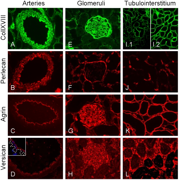
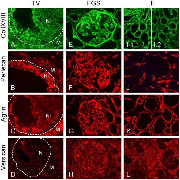
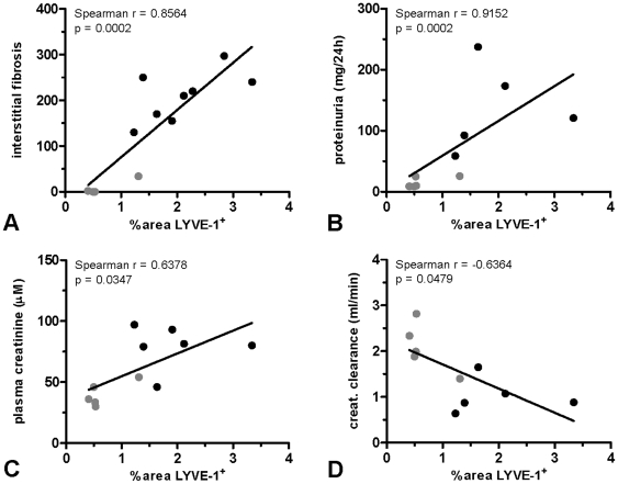
Findings: Arteries with transplant vasculopathy and sclerotic glomeruli in allografts displayed pronounced neo-expression of collXVIII and perlecan. In contrast, in interstitial fibrosis expression of the chondroitin sulfate proteoglycan versican dominated. In the cortical tubular basement membranes in both iso- and allografts, induction of collXVIII was detected. Allografts presented extensive lymphangiogenesis (p<0.01 compared to isografts and non-transplanted controls), which was associated with induced perlecan expression underneath the lymphatic endothelium (p<0.05 and p<0.01 compared to isografts and non-transplanted controls, respectively). Both the magnitude of lymphangiogenesis and perlecan expression correlated with severity of interstitial fibrosis and impaired graft function.
Interpretation: Our results reveal that changes in the extent of expression and the type of proteoglycans being expressed are tightly associated with tissue remodeling after renal transplantation. Therefore, proteoglycans might be potential targets for clinical intervention in renal chronic transplant dysfunction.
Conflict of interest statement
Figures


Similar articles
-
Renal heparan sulfate proteoglycans modulate fibroblast growth factor 2 signaling in experimental chronic transplant dysfunction.Am J Pathol. 2013 Nov;183(5):1571-1584. doi: 10.1016/j.ajpath.2013.07.030. Epub 2013 Sep 11. Am J Pathol. 2013. PMID: 24035513
-
Inflammatory lymphangiogenesis in a rat transplant model of interstitial fibrosis and tubular atrophy.Transpl Int. 2012 Jul;25(7):792-800. doi: 10.1111/j.1432-2277.2012.01482.x. Epub 2012 Apr 25. Transpl Int. 2012. PMID: 22533613
-
Chronic allograft nephropathy: expression and localization of PAI-1 and PPAR-gamma.Nephrol Dial Transplant. 2005 Dec;20(12):2812-9. doi: 10.1093/ndt/gfi172. Epub 2005 Oct 12. Nephrol Dial Transplant. 2005. PMID: 16221712
-
Restorative and rejection-associated lymphangiogenesis after renal transplantation: friend or foe?Transplantation. 2009 Dec 15;88(11):1237-9. doi: 10.1097/TP.0b013e3181c1afa7. Transplantation. 2009. PMID: 19996921 Review.
-
Proteoglycans in liver cancer.World J Gastroenterol. 2016 Jan 7;22(1):379-93. doi: 10.3748/wjg.v22.i1.379. World J Gastroenterol. 2016. PMID: 26755884 Free PMC article. Review.
Cited by
-
Predictive urinary biomarkers for steroid-resistant and steroid-sensitive focal segmental glomerulosclerosis using high resolution mass spectrometry and multivariate statistical analysis.BMC Nephrol. 2014 Sep 2;15:141. doi: 10.1186/1471-2369-15-141. BMC Nephrol. 2014. PMID: 25182141 Free PMC article.
-
Basement membrane zone collagens XV and XVIII/proteoglycans mediate leukocyte influx in renal ischemia/reperfusion.PLoS One. 2014 Sep 4;9(9):e106732. doi: 10.1371/journal.pone.0106732. eCollection 2014. PLoS One. 2014. PMID: 25188209 Free PMC article.
-
Renal interstitial fibrosis: mechanisms and evaluation.Curr Opin Nephrol Hypertens. 2012 May;21(3):289-300. doi: 10.1097/MNH.0b013e3283521cfa. Curr Opin Nephrol Hypertens. 2012. PMID: 22449945 Free PMC article. Review.
-
Selective Deletion of Heparan Sulfotransferase Enzyme, Ndst1, in Donor Endothelial and Myeloid Precursor Cells Significantly Decreases Acute Allograft Rejection.Sci Rep. 2018 Sep 7;8(1):13433. doi: 10.1038/s41598-018-31779-7. Sci Rep. 2018. PMID: 30194334 Free PMC article.
-
Differential Expression of Specific Dermatan Sulfate Domains in Renal Pathology.PLoS One. 2015 Aug 31;10(9):e0134946. doi: 10.1371/journal.pone.0134946. eCollection 2015. PLoS One. 2015. PMID: 26322947 Free PMC article.
References
-
- Kouwenhoven EA, IJzermans JN, de Bruin RW. Etiology and pathophysiology of chronic transplant dysfunction. Transpl Int. 2000;13:385–401. - PubMed
-
- Chapman JR, O'Connell PJ, Nankivell BJ. Chronic renal allograft dysfunction. J Am Soc Nephrol. 2005;16:3015–3026. - PubMed
-
- Solez K, Colvin RB, Racusen LC, Sis B, Halloran PF, et al. Banff '05 Meeting Report: differential diagnosis of chronic allograft injury and elimination of chronic allograft nephropathy (‘CAN’). Am J Transplant. 2007;7:518–526. - PubMed
-
- Solez K, Colvin RB, Racusen LC, Haas M, Sis B, et al. Banff 07 classification of renal allograft pathology: updates and future directions. Am J Transplant. 2008;8:753–760. - PubMed
-
- El Zoghby ZM, Stegall MD, Lager DJ, Kremers WK, Amer H, et al. Identifying specific causes of kidney allograft loss. Am J Transplant. 2009;9:527–535. - PubMed
Publication types
MeSH terms
Substances
LinkOut - more resources
Full Text Sources
Medical
Miscellaneous
