Depressive Symptoms and Brain Metabolite Alterations in Subjects at Ultra-high Risk for Psychosis: A Preliminary Study
- PMID: 20140124
- PMCID: PMC2808795
- DOI: 10.4306/pi.2009.6.4.264
Depressive Symptoms and Brain Metabolite Alterations in Subjects at Ultra-high Risk for Psychosis: A Preliminary Study
Abstract
Objective: Recent neuroimaging studies have suggested that brain changes occur in subjects at ultra-high risk (UHR) for psychosis while experiencing prodromal symptoms, among which depression may increase the risk of developing a psychotic disorder. The goal of this study is to examine brain metabolite levels in the anterior cingulate cortex, the left dorsolateral prefrontal cortex and the left thalamus in subjects at UHR for psychosis and to compare brain metabolite levels between the UHR subjects with comorbid major depressive disorder and healthy controls.
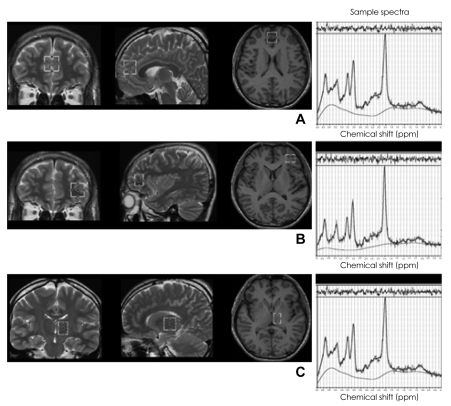
Methods: Proton magnetic resonance spectroscopy was used to examine brain metabolite levels. Twenty UHR subjects and 20 age- and intelligence quotient (IQ)-matched healthy controls were included in this study.
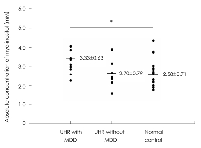
Results: Overall, no significant differences were observed in any metabolite between the UHR and healthy control group. However, UHR subjects with major depressive disorder showed significantly higher myo-inositol (Ins) levels in the left thalamus, compared to the healthy control.
Conclusion: Our results demonstrate that increased thalamic Ins level is associated with prodromal depressive symptoms. Further longitudinal follow-up studies with larger UHR sample sizes are required to investigate the function of Ins concentrations as a biomarker of vulnerability to psychosis.
Keywords: Depression; Magnetic resonance spectroscopy; Schizophrenia; Ultra-high risk.
Figures
Similar articles
-
Neural correlate of impulsivity in subjects at ultra-high risk for psychosis.Prog Neuropsychopharmacol Biol Psychiatry. 2013 Aug 1;45:165-9. doi: 10.1016/j.pnpbp.2013.04.008. Epub 2013 Apr 16. Prog Neuropsychopharmacol Biol Psychiatry. 2013. PMID: 23603064
-
Proton magnetic resonance spectroscopy in first episode psychosis and ultra high-risk individuals.Schizophr Bull. 2003;29(4):831-43. doi: 10.1093/oxfordjournals.schbul.a007049. Schizophr Bull. 2003. PMID: 14989417
-
Cerebral Glutamate and Gamma-Aminobutyric Acid Levels in Individuals at Ultra-high Risk for Psychosis and the Association With Clinical Symptoms and Cognition.Biol Psychiatry Cogn Neurosci Neuroimaging. 2020 Jun;5(6):569-579. doi: 10.1016/j.bpsc.2019.12.005. Epub 2019 Dec 19. Biol Psychiatry Cogn Neurosci Neuroimaging. 2020. PMID: 32008981
-
Brain structural abnormalities as potential markers for detecting individuals with ultra-high risk for psychosis: A systematic review and meta-analysis.Schizophr Res. 2019 Jul;209:22-31. doi: 10.1016/j.schres.2019.05.015. Epub 2019 May 16. Schizophr Res. 2019. PMID: 31104914
-
[Stress and psychotic transition: A literature review].Encephale. 2016 Aug;42(4):367-73. doi: 10.1016/j.encep.2015.10.001. Epub 2016 May 6. Encephale. 2016. PMID: 27161263 Review. French.
Cited by
-
N-Acetylaspartate and Choline Metabolites in Cortical and Subcortical Regions in Clinical High Risk Relative to Healthy Control Subjects: An Exploratory 7T MRSI Study.Int J Mol Sci. 2023 Apr 22;24(9):7682. doi: 10.3390/ijms24097682. Int J Mol Sci. 2023. PMID: 37175389 Free PMC article.
-
Niacin Skin Sensitivity Is Increased in Adolescents at Ultra-High Risk for Psychosis.PLoS One. 2016 Feb 19;11(2):e0148429. doi: 10.1371/journal.pone.0148429. eCollection 2016. PLoS One. 2016. PMID: 26894921 Free PMC article.
-
Dorsolateral Prefrontal Cortex Glutamate/Gamma-Aminobutyric Acid (GABA) Alterations in Clinical High Risk and First-Episode Schizophrenia: A Preliminary 7-T Magnetic Resonance Spectroscopy Imaging Study.Int J Mol Sci. 2022 Dec 13;23(24):15846. doi: 10.3390/ijms232415846. Int J Mol Sci. 2022. PMID: 36555487 Free PMC article.
-
Abnormal metabolites in the dorsolateral prefrontal cortex of female epilepsy patients with migraine without aura.Neuroreport. 2024 Dec 11;35(18):1155-1162. doi: 10.1097/WNR.0000000000002110. Epub 2024 Nov 6. Neuroreport. 2024. PMID: 39526657 Free PMC article.
-
Dopamine and glutamate in individuals at high risk for psychosis: a meta-analysis of in vivo imaging findings and their variability compared to controls.World Psychiatry. 2021 Oct;20(3):405-416. doi: 10.1002/wps.20893. World Psychiatry. 2021. PMID: 34505389 Free PMC article.
References
-
- Borgwardt SJ, Riecher-Rössler A, Dazzan P, Chitnis X, Aston J, Drewe M, et al. Regional gray matter volume abnormalities in the at risk mental state. Biol Psychiatry. 2007;61:1148–1156. - PubMed
-
- Velakoulis D, Wood SJ, Wong MT, McGorry PD, Yung A, Phillips L, et al. Hippocampal and amygdala volumes according to psychosis stage and diagnosis: a magnetic resonance imaging study of chronic schizophrenia, first-episode psychosis, and ultra-high-risk individuals. Arch Gen Psychiatry. 2006;63:139–149. - PubMed
-
- Yücel M, Wood SJ, Phillips LJ, Stuart GW, Smith DJ, Yung A, et al. Morphology of the anterior cingulate cortex in young men at ultra-high risk of developing a psychotic illness. Br J Psychiatry. 2003;182:518–524. - PubMed
-
- Wood SJ, Berger G, Velakoulis D, Phillips LJ, McGorry PD, Yung AR, et al. Proton magnetic resonance spectroscopy in first episode psychosis and ultra high-risk individuals. Schizophr Bull. 2003;29:831–843. - PubMed
-
- Jessen F, Scherk H, Träber F, Theyson S, Berning J, Tepest R, et al. Proton magnetic resonance spectroscopy in subjects at risk for schizophrenia. Schizophr Res. 2006;87:81–88. - PubMed
LinkOut - more resources
Full Text Sources