. 2010 Feb 5;6(2):e1000754.
doi: 10.1371/journal.ppat.1000754.

TgMORN1 is a key organizer for the basal complex of Toxoplasma gondii

Affiliations

TgMORN1 is a key organizer for the basal complex of Toxoplasma gondii

Aoife T Heaslip et al. PLoS Pathog. .

Abstract

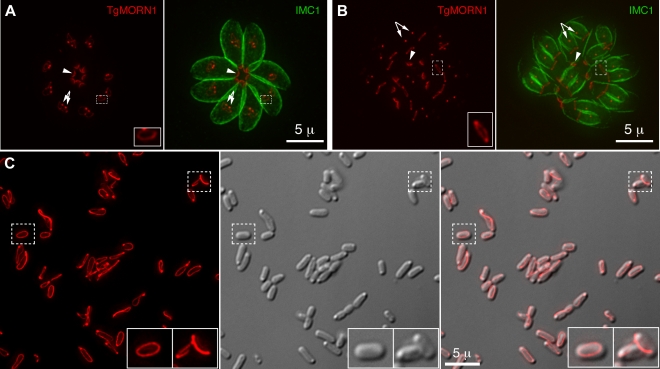
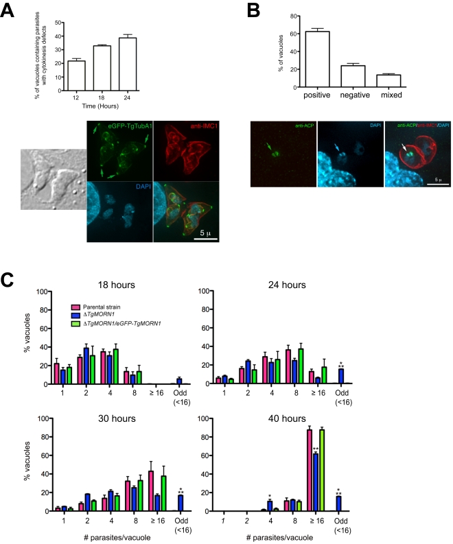
Toxoplasma gondii is a leading cause of congenital birth defects, as well as a cause for ocular and neurological diseases in humans. Its cytoskeleton is essential for parasite replication and invasion and contains many unique structures that are potential drug targets. Therefore, the biogenesis of the cytoskeletal structure of T. gondii is not only important for its pathogenesis, but also of interest to cell biology in general. Previously, we and others identified a new T. gondii cytoskeletal protein, TgMORN1, which is recruited to the basal complex at the very beginning of daughter formation. However, its function remained largely unknown. In this study, we generated a knock-out mutant of TgMORN1 (DeltaTgMORN1) using a Cre-LoxP based approach. We found that the structure of the basal complex was grossly affected in DeltaTgMORN1 parasites, which also displayed defects in cytokinesis. Moreover, DeltaTgMORN1 parasites showed significant growth impairment in vitro, and this translated into greatly attenuated virulence in mice. Therefore, our results demonstrate that TgMORN1 is required for maintaining the structural integrity of the parasite posterior end, and provide direct evidence that cytoskeleton integrity is essential for parasite virulence and pathogenesis.


Conflict of interest statement

The authors have declared that no competing interests exist.

Figures

Figure 1
Figure 1. TgMORN1 is a component of the basal complex and forms rings and fibers when ectopically expressed in E. coli.
A–B. The localization of TgMORN1 in parasites at the beginning (A) or late stage (B) of cell division. Red: eGFP-TgMORN1 pseudocolored red. Green: anti-IMC1 antibody labeling highlighting the protein network underneath the inner membrane complex (IMC). Arrowheads: the basal complexes of mother parasites. Arrows: spindle poles. Insets indicate the basal ring complexes of daughters and are at 2.5× magnification. C. 6XHIS-mCherryFP-TgMORN1 (red) expressed in E. coli formed rings and fibers. Left: Fluorescent image of mCherryFP-TgMORN1 containing rings and fibers. Middle: DIC image. Right: Overlay of the fluorescent image and the DIC. Insets are at 2× magnification.
Figure 2
Figure 2. The generation of TgMORN1 knockout (ΔTgMORN1) parasites.
A. Diagram describing the procedure for generating the ΔTgMORN1 parasite line. See text for details. B. Genomic PCR analysis of RHΔHX, the parental (LoxP-TgMORN1-HXGPRT-LoxP), and ΔTgMORN1 parasites. The diagram at the top shows the positions of the sequences that the PCR primers (A1, A2, S1, S2, S3) hybridize with. Boxes marked by slanted lines indicate regions of 5′ and 3′ UTR of TgMORN1 gene included in LoxP_TgMORN1_HXGPRT_LoxP knock-in plasmid (c.f. Figure 2A ). C. Western blot analysis of RHΔHX, parental, ΔTgMORN1, and the complemented (ΔTgMORN1/eGFP-TgMORN1) parasites, which shows that the level of TgMORN1 expression was comparable among RHΔHX, the parental strain and the complemented parasites, but was undetectable in ΔTgMORN1 parasites. The blot was reprobed with mouse-anti-tubulin B-5-1-2 to use α-tubulin as a loading control.
Figure 3
Figure 3. The loss of TgMORN1 affects the organization of the parasite posterior end.
A. The comparison of the distribution of the width of basal IMC1 gap in the parental strain (n = 102), ΔTgMORN1 (n = 114), and the complemented - ΔTgMORN1/eGFP-TgMORN1 parasites (n = 118). Red lines indicate the average. B. The comparison of parasite morphology of the parental strain (top panels), ΔTgMORN1 (middle panels), and the complemented (bottom panels) parasites in vacuoles containing one or two parasites. Notice the heterogeneity of the posterior end morphology in ΔTgMORN1 parasites. Three main classes of morphology are shown: parasites with wide basal IMC1 gap (white arrowheads), parasites with small or no basal IMC1 gap but wide posterior end (white arrow), and parasites with a largely normal posterior end that sometimes contained irregular IMC structure (purple arrow). Green: anti-IMC1 antibody labeling. Red: anti-TgMORN1 labeling (the parental strain) or eGFP-TgMORN1 labeling (the complement). C. The comparison of parasite morphology of the parental strain (top panels), ΔTgMORN1 (middle panels), and the complemented (bottom panels) parasites in vacuoles ≥16 parasites. Notice the highly irregular organization of ΔTgMORN1 parasites within the vacuole. Green: anti-IMC1 antibody labeling. Red: anti-TgMORN1 labeling (the parental strain, ΔTgMORN1 parasites) or eGFP-TgMORN1 labeling (the complement).
Figure 4
Figure 4. The loss of TgMORN1 affects the localization of other proteins to the basal complex.
A. When eGFP-TgCentrin2 (pseudo-colored red) was expressed in ΔTgMORN1 parasites (bottom panels), it was incorporated into the apical complex, the centrioles and the peripheral annuli (arrows) as in the parental strain (top panels), but eGFP-TgCentrin2 basal complex localization (arrowheads, insets) was undetectable in these parasites. Insets are at 2× magnification and contrast enhanced to visualize the basal eGFP-TgCentrin2 structure. B. When eGFP-TgDLC (pseudo-colored red) was expressed in ΔTgMORN1 parasites (bottom panels), it was incorporated into the apical complex, and spindle pole/ centriole assembly (red arrows) as in the parental strain (top panels), but an eGFP-TgDLC concentration could not be detected in the parasite posterior end (red arrowheads, insets). White arrows indicate the apical complexes of two parasites that failed to complete cytokinesis in the previous round of replication. Insets are at 2× magnification and contrast enhanced to visualize the basal eGFP-TgDLC structure.
Figure 5
Figure 5. The loss of TgMORN1 has little effect in invasion.
Graph shows the number of parasites invaded after equal number of extracellular parental, ΔTgMORN1 or ΔTgMORN1/eGFP-TgMORN1 parasites were added to confluent HFF monolayers and incubated at 37°C for 1 hour. Result is the summary of three independent experiments. Error bar: standard error of the mean. P values for the comparison between ΔTgMORN1 and parental parasites; and ΔTgMORN1 and complemented parasites are 0.27 and 0.35 respectively (Student t-test).
Figure 6
Figure 6. ΔTgMORN1 parasites display defects in cytokinesis and apicoplast segregation.
A. Top: Graph shows the percentages of vacuoles with ΔTgMORN1 parasites displaying cytokinesis defects at 12, 18, 24 hours post infection. Error bar: standard error of the mean. Bottom: Images show a parasitophorous vacuole containing two pairs of ΔTgMORN1 parasites, both of which have failed cytokinesis in the previous round of cell division. These parasites expressed eGFP-TgTubA1 (green) and were also labeled with anti-IMC1 (red), showing that the apical complex, cortical microtubules and IMC1 network all formed but the parasites failed to separate before beginning the next round of daughter construction. Arrows indicate the apical complexes of the mother parasites. B. Top: Graph shows the percentages of vacuoles with ΔTgMORN1 parasites scored as apicoplast positive (i.e. all parasites in the vacuole contain apicoplast, ∼62%), apicoplast negative (i.e. none of the parasites in the vacuole contains apicoplast, ∼24%) or mixed (i.e. vacuoles contains both apicoplast positive and negative parasites, ∼14%) (n = 250). Error bar: standard error of the mean. No apicoplast negative parasites were observed in either the parental or complemented strains (n = 50). Bottom: Images show a parasitophorous vacuole containing two ΔTgMORN1 parasites, where one contains apicoplast (arrows) and the other one does not. Green: anti-ACP; red: anti-IMC1; cyan: DAPI. C. Comparison of the intracellular growth of ΔTgMORN1 parasites with those of the parental strain and ΔTgMORN1/eGFP-TgMORN1 parasites 18, 24, 30 and 40 hours after infection. The trend shows a decrease in replication rate upon the loss of TgMORN1. It also shows that asynchronized replication within the parasitophorous vacuole occurred much more frequently in ΔTgMORN1 parasites than in the parental strain and ΔTgMORN1/eGFP-TgMORN1 parasites. “Odd” group includes vacuoles in which the number of parasites was less than 16, and not an integral power of 2. “≥16” group includes all the vacuoles that contained 16 or more parasites. Error bar: standard error of the mean. “*” indicates P values less than 0.05, “**” indicates P values less than 0.01, and “***” indicates P values less than 0.0001 (Student t-test), when ΔTgMORN1 parasites are compared with the parental strain or ΔTgMORN1/eGFP-TgMORN1 parasites. For the 40-hour time point, vacuoles containing one and two parasites were not included in the counting, as they were most likely secondary vacuoles formed by parasites that egressed from large primary vacuoles and reinvaded.
Figure 7
Figure 7. ΔTgMORN1 parasites display growth defects in vitro and greatly attenuated virulence in mice.
A. ΔTgMORN1 parasites formed plaques (middle, arrow) significantly smaller than those formed by the parental (left), and the complemented (ΔTgMORN1/eGFP-TgMORN1) strains (right) (arrowheads). Insets are at 2× magnification. B. CD1 outbred mice were infected intraperitoneally with either 103 or 104 parental; 103, 104, 2×10⁴ or 105 ΔTgMORN1; or 103 or 104 ΔTgMORN1/eGFP-TgMORN1 parasites. Mice that were infected with parental parasites died between day 7 and day 9 pi. In contrast, mice that were challenged with 103, 104, 2×10⁴ or 105 ΔTgMORN1 parasites remained alive. EGFP-TgMORN1 complementation restored parasite virulence. For the parental and ΔTgMORN1/eGFP-TgMORN1 parasites, only the data for 103 parasites infection is shown. All surviving mice remain healthy for more than 60 days after infection. Data for only the first 30 days of the experiment is shown. C. At day 21 pi, surviving mice “immunized” with 103 or 104 ΔTgMORN1 parasites were challenged with 10,000 wild type RH parasites (LD100 = 1). Compared to naïve mice, which died between day 7 and day 10 pi, mice that were pre-infected with ΔTgMORN1 parasites were protected from lethal challenge, where all mice immunized with 104 ΔTgMORN1 parasites and 75% of mice immunized with 103 ΔTgMORN1 parasites remained alive. All surviving mice remain healthy for more than 60 days after the challenge. Data for only the first 30 days of the experiment is shown.

Similar articles

Cited by

References

    1. Levine ND. Progress in taxonomy of the Apicomplexan protozoa. J Protozool. 1988;35:518–520. - PubMed
    1. Zuckerman A. Recent studies on factors involved in malarial anemia. Military Medicine. 1966;131(Suppl):1201–1216. - PubMed
    1. Nagel RL. Malarial anemia. Hemoglobin. 2002;26:329–343. - PubMed
    1. Ehrhardt S, Burchard GD, Mantel C, Cramer JP, Kaiser S, et al. Malaria, anemia, and malnutrition in african children–defining intervention priorities. Journal of Infectious Diseases. 2006;194:108–114. - PubMed
    1. Nichols BA, Chiappino ML. Cytoskeleton of Toxoplasma gondii. J Protozool. 1987;34:217–226. - PubMed

Publication types