. 2010 Feb 5;6(2):e1000755.
doi: 10.1371/journal.ppat.1000755.

Neutrophil-derived CCL3 is essential for the rapid recruitment of dendritic cells to the site of Leishmania major inoculation in resistant mice

Affiliations

Neutrophil-derived CCL3 is essential for the rapid recruitment of dendritic cells to the site of Leishmania major inoculation in resistant mice

Mélanie Charmoy et al. PLoS Pathog. .

Abstract

Neutrophils are rapidly and massively recruited to sites of microbial infection, where they can influence the recruitment of dendritic cells. Here, we have analyzed the role of neutrophil released chemokines in the early recruitment of dendritic cells (DCs) in an experimental model of Leishmania major infection. We show in vitro, as well as during infection, that the parasite induced the expression of CCL3 selectively in neutrophils from L. major resistant mice. Neutrophil-secreted CCL3 was critical in chemotaxis of immature DCs, an effect lost upon CCL3 neutralisation. Depletion of neutrophils prior to infection, as well as pharmacological or genetic inhibition of CCL3, resulted in a significant decrease in DC recruitment at the site of parasite inoculation. Decreased DC recruitment in CCL3(-/-) mice was corrected by the transfer of wild type neutrophils at the time of infection. The early release of CCL3 by neutrophils was further shown to have a transient impact on the development of a protective immune response. Altogether, we identified a novel role for neutrophil-secreted CCL3 in the first wave of DC recruitment to the site of infection with L. major, suggesting that the selective release of neutrophil-secreted chemokines may regulate the development of immune response to pathogens.


Conflict of interest statement

AEIP is an employee of Merck-Serono Research center in Geneva. All of the other authors declare no conflict of interest.

Figures
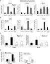
Figure 1
Figure 1. In presence of L. major, neutrophils produce CCL3 which chemo-attracts bone marrow-derived DCs in vitro.
L. major–i.p. recruited C57BL/6 and BALB/c neutrophils were purified by MACS and incubated with medium, L. major (5∶1 parasite∶neutrophil ratio), IFNγ, or both. (A) Sixteen hours later, neutrophil CCL3, CCL4 and CCL5 mRNA levels were assessed by real-time quantitative RT-PCR, and (B) 24 hours after initiation of culture, chemokine content was measured in cell-free culture supernatants by ELISA. Data are the mean triplicate measurement ± SEM of neutrophil mRNA or chemokine content in the supernatants. *: p<0.05 compared to neutrophils cultured with medium only. (C) Supernatants from inflammatory neutrophils cultured in presence or in absence of L. major were tested for their chemotactic activity towards bone marrow-derived DCs in a transwell cell migration assay. The number of DCs that migrated toward neutrophil supernatant is represented. (D) DC migration was similarly assessed in response to neutrophil supernatants depleted of CCL3. *: p<0.05 as compared to values measured in response to supernatant of neutrophil incubated with medium alone. Data are expressed as mean number ± SEM of DC that migrated towards neutrophil supernatant (n = 3 per group). The results of a representative experiment out of three are shown.
Figure 2
Figure 2. Higher number of DCs are recruited to the ear dermis of L. major infected C57BL/6 compared to BALB/c mice.
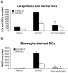
(A) The number of neutrophils, Langerhans and dermal DCs, and monocyte-derived DCs emigrating from ear explants was measured at 0, 6, 24 and 48 hours post L. major inoculation *: p<0.05 comparing cell number in C57BL/6 versus BALB/c mice. Values from ear explants of six mice per group are expressed as mean values ± SEM . The data are representative of three separate experiments. (B) Gating strategy by flow cytometry of Langerhans and dermal DCs, and monocyte-derived DCs emigrating from L. major-infected C57BL/6 ear explants. For Langerhans and dermal DCs, MHCII and DEC205 positive cells were analyzed on CD11c+ gated cells. For monocyte-derived DCs, four colour FACS analysis was performed, CD11b+ and LY6C+ cells were analyzed on a CCD11cdim and Ly6G gated cell population. A representative flow cytometry plot is shown.
Figure 3
Figure 3. Neutrophils are essential for DC recruitment to the ear dermis following L. major promastigote inoculation.
C57BL/6 and BALB/c mice were depleted of neutrophils by an injection i.p. of the NIMP-R14 anti-neutrophil mAb, or injected with a control mAb, 6 hours prior to infection i.d. with L. major stationary promastigotes. The number of (A) Langerhans and dermal DCs and (B) monocyte-derived DCs recruited in the ear dermis at 0h (naïve) or 24h following L. major inoculation was quantified and compared in mice depleted or not of neutrophils. Data are given as mean values ± SEM (n = 6 per group) and are representative of three experiments. * : p<0.05 between mice depleted or not of neutrophils.
Figure 4
Figure 4. CCL3 is a major DC-attracting chemokine in the ear dermis one day post L. major inoculation.
(A) C57BL/6 and BALB/c mice were infected with L. major in the ear dermis. mRNA expression of CCL3, CCL4 and CCL5 at the site of infection was measured 0, 6, 24 and 48 hours post infection by quantitative Real-time PCR and normalized relative to HPRT mRNA levels. Data are represented as the mean ± SEM mRNA transcript levels of individual infected ear (n = 6 per time point). One representative experiment of three is shown. (B) Twenty four hours post-infection, the ear chemokine mRNA level was compared in C57BL/6 mice that were given either the NIMP-R14 neutrophil depleting mAb or a control mAb 6 hours prior to L. major inoculation in the ear. Results are represented as fold increase in mRNA levels relative to levels measured in uninfected mice given a value of 1, and are representative of two experiments.
Figure 5
Figure 5. CCL3 is essential for early DC trafficking/recruitment to the site of infection following L. major inoculation.
(A) C57BL/6 and BALB/c mice were injected i.p. with Evasin-1, a chemokine binding protein that neutralizes CCL3. Twenty four hours post L. major inoculation, the number of neutrophils, Langerhans and dermal DCs, and monocyte-derived DCs, spontaneously emigrating out of ear explants was measured and compared to that obtained from ear explants from mice similarly infected but which were not given Evasin-1. * : p<0.05 between mice treated or not with Evasin-1. (B) 24 hours after infection i.d. with L. major, the number of leukocytes emigrating out of ear skin explants of CCL3−/− mice was compared to that measured in similarly infected C57BL/6 ear explants. The number of neutrophils, Langerhans and dermal DCs, and monocyte-derived DCs is presented as the mean ± SEM (n = 4 for Evasin-1 treated mice, and n = 6 for CCL3−/− mice) and are representative of three experiments. *: p<0.05 between CCL3−/− and C57BL/6 mice.
Figure 6
Figure 6. The CCL3 secreted by neutrophils is the major chemoattractant for DCs one day after L. major inoculation.
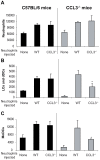
Inflammatory neutrophils from C57BL/6 or CCL3−/− mice were injected in the ear dermis of C57BL/6 or CCL3−/− mice simultaneously with L. major. (A) The neutrophils, (B) Langerhans cells, dermal DCs, and (C) monocyte-derived DCs emigrating from the ear explants were monitored 24 hours post inoculation. Data obtained from four individual mice are expressed as mean values ±SEM. The results are representative of three experiments. * : p<0.05 compared with injection with WT neutrophils.
Figure 7
Figure 7. Depletion of CCL3 during the first day post L. major inoculation delays the development of the L. major-induced Th1 immune response.
CCL3−/− mice, and C57BL/6 mice in which CCL3 was blocked by injection of Evasin-1 for the first five days post L. major inoculation, were inoculated s.c. with 3×10⁶ L. major. CD4+ draining lymph nodes cells were prepared at days 15 (A) and 40 (B) post L. major inoculation and co-incubated with UV irradiated L. major. The resulting supernatants were monitored for their IFNγ and IL-4 content by ELISA. Data are the mean of triplicate measurement ± SEM of cytokines (n = 8 mice per group). Fifteen (C) or forty (D) days post-infection L. major-specific IgG2a and IgG1 Ab production was quantified in sera from infected mice. Data are the mean triplicate OD ± SEM of serum dilution. (E) Course of infection in CCL3−/− and Evasin-1 treated mice. Evolution of lesion size was monitored every week with a Vernier caliper. Each point represents the mean lesion size ±SEM (n = 8 per group). Parasite burden was measured by limiting dilution assay (LDA) fifteen (F) and forty (G) days post inoculation (n = 8 per group). The experiments are representative of five (15 days) and three (6 weeks) independent experiments.

References

    1. Nathan C. Neutrophils and immunity: challenges and opportunities. Nat Rev Immunol. 2006;6:173–182. - PubMed
    1. Appelberg R. Neutrophils and intracellular pathogens: beyond phagocytosis and killing. Trends Microbiol. 2007;15:87–92. - PubMed
    1. Sacks D, Noben-Trauth N. The immunology of susceptibility and resistance to Leishmania major in mice. Nat Rev Immunol. 2002;2:845–858. - PubMed
    1. Beil WJ, Meinardus-Hager G, Neugebauer DC, Sorg C. Differences in the onset of the inflammatory response to cutaneous leishmaniasis in resistant and susceptible mice. J Leukoc Biol. 1992;52:135–142. - PubMed
    1. Tacchini-Cottier F, Zweifel C, Belkaid Y, Mukankundiye C, Vasei M, et al. An immunomodulatory function for neutrophils during the induction of a CD4+ Th2 response in BALB/c mice infected with Leishmania major. J Immunol. 2000;165:2628–2636. - PubMed

Publication types

LinkOut - more resources