. 2010 Feb 5;6(2):e1000756.
doi: 10.1371/journal.ppat.1000756.

Exacerbated innate host response to SARS-CoV in aged non-human primates

Affiliations

Exacerbated innate host response to SARS-CoV in aged non-human primates

Saskia L Smits et al. PLoS Pathog. .

Abstract

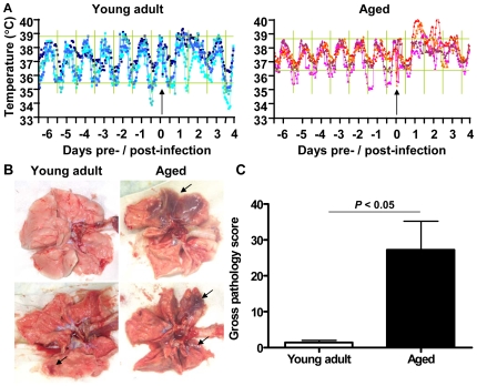
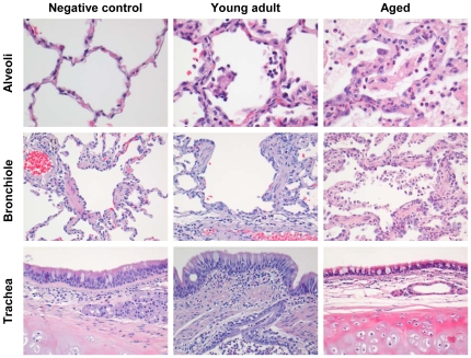
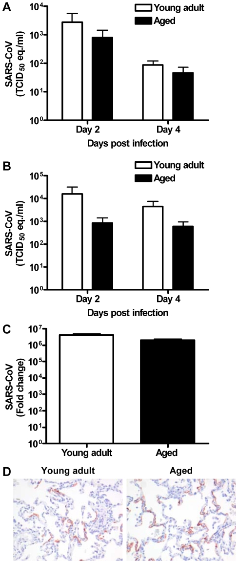
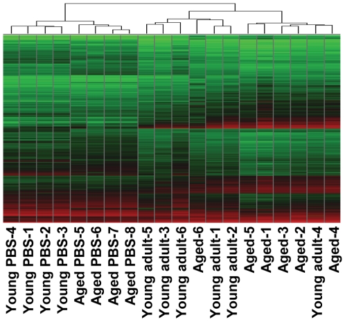
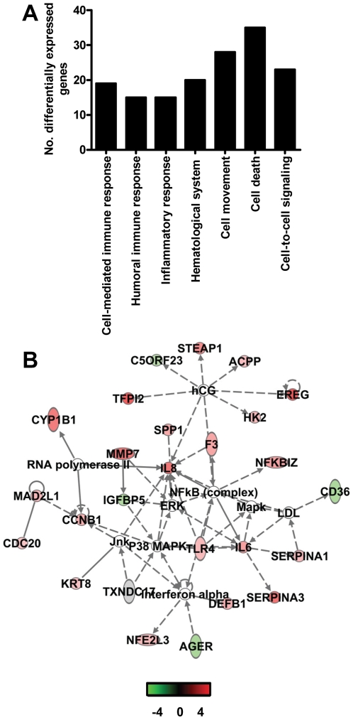
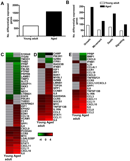
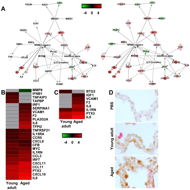
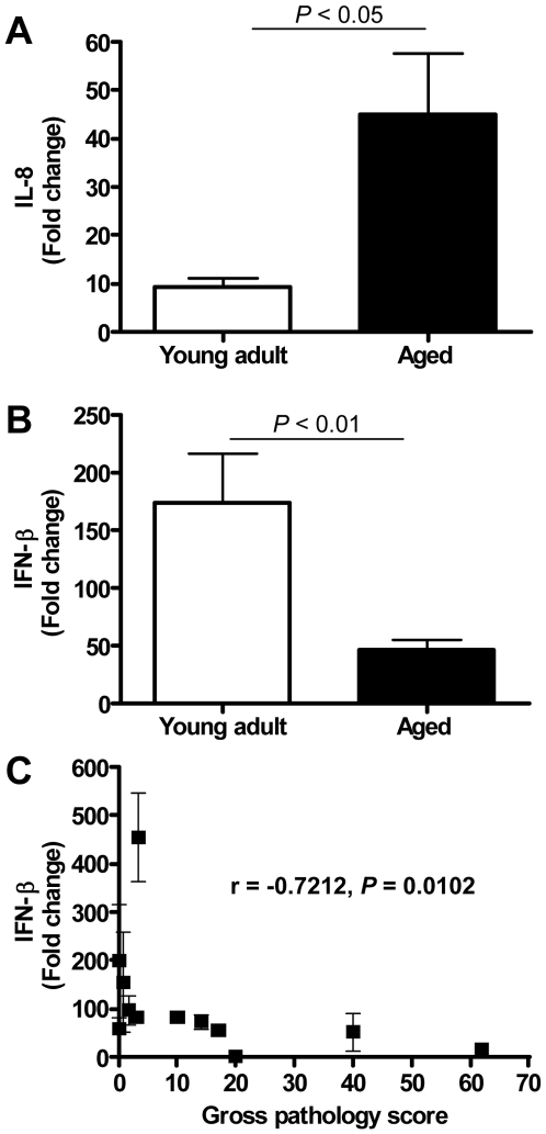
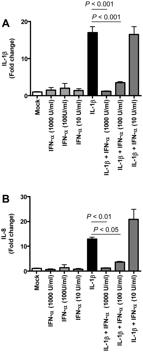
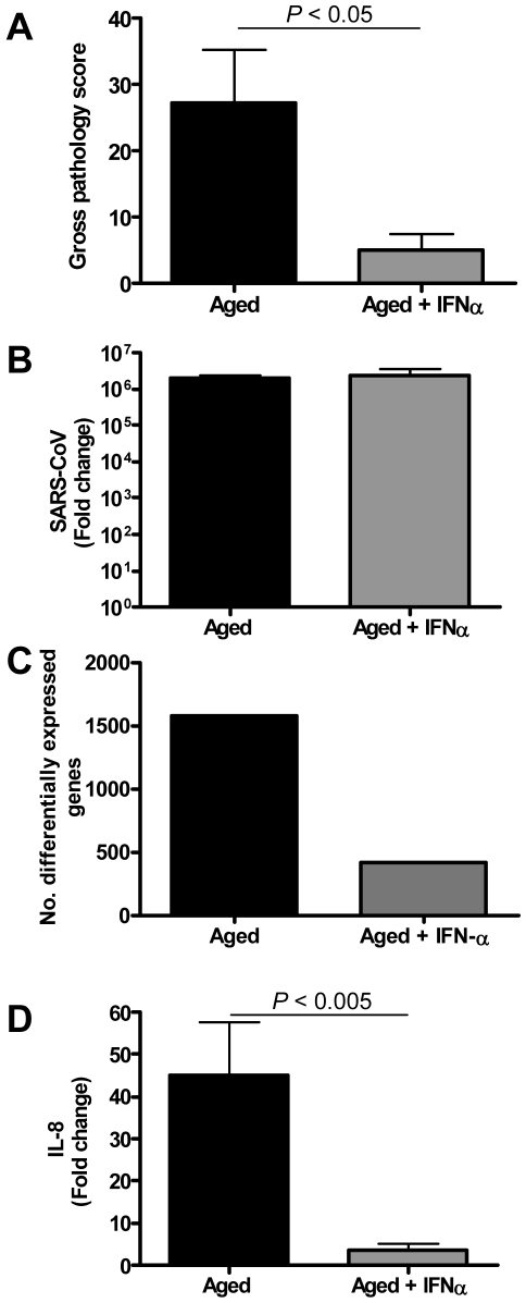
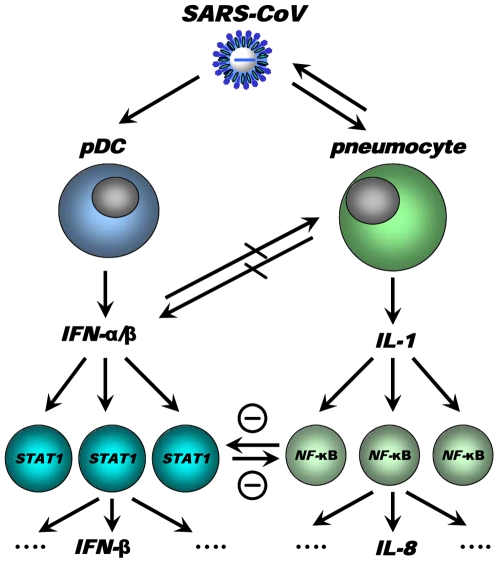
The emergence of viral respiratory pathogens with pandemic potential, such as severe acute respiratory syndrome coronavirus (SARS-CoV) and influenza A H5N1, urges the need for deciphering their pathogenesis to develop new intervention strategies. SARS-CoV infection causes acute lung injury (ALI) that may develop into life-threatening acute respiratory distress syndrome (ARDS) with advanced age correlating positively with adverse disease outcome. The molecular pathways, however, that cause virus-induced ALI/ARDS in aged individuals are ill-defined. Here, we show that SARS-CoV-infected aged macaques develop more severe pathology than young adult animals, even though viral replication levels are similar. Comprehensive genomic analyses indicate that aged macaques have a stronger host response to virus infection than young adult macaques, with an increase in differential expression of genes associated with inflammation, with NF-kappaB as central player, whereas expression of type I interferon (IFN)-beta is reduced. Therapeutic treatment of SARS-CoV-infected aged macaques with type I IFN reduces pathology and diminishes pro-inflammatory gene expression, including interleukin-8 (IL-8) levels, without affecting virus replication in the lungs. Thus, ALI in SARS-CoV-infected aged macaques developed as a result of an exacerbated innate host response. The anti-inflammatory action of type I IFN reveals a potential intervention strategy for virus-induced ALI.


Conflict of interest statement

The authors have declared that no competing interests exist.

Figures

Figure 1
Figure 1. Aged macaques are more prone to develop SARS-CoV-associated disease than young adults.
(A) Fluctuations in body temperatures in four young adult and four aged SARS-CoV-infected macaques measured by transponders in the peritoneal cavity. Temperatures are shown from day six prior to infection until four days post infection. The arrow indicates day zero when animals were infected. Grey horizontal lines mark the average range of temperature fluctuations prior to infection. (B) Macroscopic appearance of (consolidated) lung tissue of young adult and aged SARS-CoV infected macaques at day 4 post infection. Lesions are arrowed. (C) Gross pathology scores of aged and young adult macaque groups were determined after necropsy and averaged (±standard error of the mean (s.e.m.)).
Figure 2
Figure 2. Histology of lungs from SARS-CoV-infected aged macaques.
Lesions in lungs of PBS-infected (left panel) and SARS-CoV infected young adult (middle panel) and aged (right panel) macaques showing diffuse alveolar damage, characterized by disruption of alveolar walls causing edema and type II pneumocyte hyperplasia with influx of inflammatory cells in the alveoli and bronchioles. In the trachea, a multifocal mild chronic lymphoplasmacytic tracheobronchoadenitis was observed in young adult macaques.
Figure 3
Figure 3. Viral replication levels in SARS-CoV-infected aged and young adult macaques are similar.
(A–B) SARS-CoV replication in the throat (A) and nose (B) of SARS-CoV infected aged (black bars) and young adult (white bars) macaques at day 2 and 4 post infection as determined by real-time RT-PCR. Viral RNA levels are displayed as TCID50 equivalents (eq.)/ml swab medium (±s.e.m.). (C) Average fold change in SARS-CoV mRNA levels (±s.e.m.) in the lungs of aged and young adult macaques compared to PBS-infected animals as determined by real-time RT-PCR and depicted on a log-scale. (D) Lung sections of SARS-CoV-infected aged and young adult macaques were stained with a mouse-anti-SARS-nucleocapsid IgG2a. Sections were counterstained with hematoxylin. Original magnifications are ×20.
Figure 4
Figure 4. Global gene expression profiles of individual young adult and aged animals.
Global gene expression profiles of normalized log-2 based hybridization signals of individual young adult and aged macaques of a set of gene transcripts that were identified as being differentially regulated (fold change ≥2; FDR<0.05) in at least one of the comparisons of gene expression in the lungs of experimentally SARS-CoV-infected aged and young adult macaques versus gene expression in the lungs of PBS-infected macaques, and genes were included if they met the criteria of an absolute fold change of ≥2-fold (FDR<0.05) in at least one experiment. The data were plotted as a heat map, where each matrix entry represents a gene expression value. Normalized log-2 based hybridization signals ranged from 3 (green) to 14 (red). Dendograms (trees) of the heat map represent the degree of relatedness between the samples, with short branches denoting a high degree of similarity and long branches denoting a low degree of similarity.
Figure 5
Figure 5. Direct comparison of gene expression profiles in the lungs of aged and young adult SARS-CoV-infected macaques.
(A) Number of differentially expressed genes in the direct contrast of aged and young adult SARS-CoV-infected macaques with functions in immune response, inflammatory response, hematological system development and function, cell movement, cell death, or cell-to-cell signaling and interaction obtained from Ingenuity Pathways Knowledge Base. (B) This diagram shows a gene interaction network from Ingenuity Pathways Knowledge Base with genes that are differentially expressed in the contrast of aged and young adult SARS-CoV-infected animals. The central node is NF-κB, a key transcription factor in inflammation and ARDS. Genes depicted in green are downregulated and in red upregulated.
Figure 6
Figure 6. Aged macaques display a stronger host response to SARS-CoV infection than young adults.
(A) Number of differentially expressed gene transcripts in aged and young adult SARS-CoV-infected macaques compared to aged and young adult PBS-infected animals, respectively (≥2-fold change, FDR<0.05, LIMMA analysis). (B) Number of differentially expressed genes in aged and young adult macaque groups compared to aged and young adult PBS-infected animals, respectively, with functions in cellular growth and proliferation, cell movement, cell death, or cell-to-cell signaling and interaction obtained from Ingenuity Pathways Knowledge Base. White bars for young adult and black bars for aged macaques. When SARS-CoV-infected aged and young adult macaques were compared directly, these cellular functions were significantly differentially expressed as well. (C–E) Gene expression profiles showing differentially expressed genes coding for proteins involved in cell adhesion (C), apopotosis (D), and cytokine/chemokine signalling (E) of aged and young adult macaques. Gene sets were obtained from Ingenuity Pathways Knowledge Base and changed ≥2-fold in at least one of the macaque groups as compared to PBS-infected controls. The data presented are error-weighted fold change averages for six young adult and aged animals. Genes shown in red were upregulated, in green downregulated, and in grey not significantly diferentially expressed in infected animals relative to PBS-infected animals (log (base 2) transformed expression values with minimum and maximum values of the color range being −4 and 4). Global test analysis of the direct contrast of SARS-CoV-infected aged versus young adult animals showed that these pathways were significantly differentially expressed (P<0.05). See Table S2 and S3 for full gene names and expression values.
Figure 7
Figure 7. NF-κB-signalling in aged and young adult macaques.
(A) These diagrams show a gene interaction network from Ingenuity Pathways Knowledge Base with genes that are differentially expressed in the contrast of aged SARS-CoV-infected animals versus aged PBS-infected macaques. The central node is NF-κB, a key factor in inflammation and development of ARDS. Genes depicted in green are downregulated and in red upregulated. As a reference, the same network is shown for young adult animals (left panel) and aged animals (right panel). (B–C) Gene expression profiles showing differentially expressed NF-κB target genes (B) and genes coding for proteins involved ARDS (C) of aged and young adult macaques. Gene sets were obtained from Ingenuity Pathways Knowledge Base or literature and changed ≥2-fold in at least one of the macaque groups as compared to PBS-infected controls. The data presented are error-weighted fold change averages for six young adult and aged animals. Genes shown in red were upregulated, in green downregulated, and in grey not significantly diferentially expressed in infected animals relative to PBS-infected animals (log (base 2) transformed expression values with minimum and maximum values of the color range being −4 and 4). See Table S2 and S3 for full gene names and expression values. (D) Lung sections of PBS and SARS-CoV-infected aged and young adult macaques were stained with an antibody against phosphorylated NF-κB (brown) and with a mouse-anti-SARS-nucleocapsid IgG2a (red). Sections were counterstained with hematoxylin. Original magnifications are ×40.
Figure 8
Figure 8. Quantitative RT-PCR confirmation of IFN-β mRNA levels.
(A) Quantitative RT-PCR for IL-8 was performed on two-three separate lung samples per animal with substantial virus replication. The data presented are error-weighted (±s.e.m.) averages of the fold-change as compared to PBS-infected controls for young adult (n = 6) and aged (n = 6) animals. (B) Quantitative RT-PCR for IFN-β was performed on two-three separate lung samples per animal with substantial virus replication. The data presented are error-weighted (±s.e.m.) averages of the fold-change as compared to PBS-infected controls for young adult (n = 6) and aged (n = 6) animals. (C) The expression level of IFN-β (fold change) per animal was plotted against gross pathology score and the correlation coefficient was determined using Spearman's correlation test.
Figure 9
Figure 9. Anti-inflammatory type I IFN inhibits IL-1β-induced pro-inflammatory cytokine production in PBMCs.
(A–B) Induction of IL-1β (A) and IL-8 (B) mRNAs after treatment of human PBMC with IL-1β (5 ng/ml), IFN-α (1000 U/ml, 100U/ml, or 10 U/ml) or both as determined by quantitative RT-PCR. The data presented are error-weighted (±s.e.m.) averages of the fold-change as compared to untreated (Mock) PBMC. Shown are representative data from one out of four donors.
Figure 10
Figure 10. Anti-inflammatory type I IFN inhibits virus-induced ALI in aged SARS-CoV-infected macaques.
(A) Gross pathology scores of lungs from macaques were determined during necropsy and averaged (±s.e.m.). (B) Average fold change (±s.e.m.) in SARS-CoV mRNA levels in the lungs of pegylated IFN-α-treated (n = 3) and untreated aged (n = 6) macaques compared to aged PBS-infected (n = 4) animals as determined by real-time RT-PCR. (C) Number of differentially expressed gene transcripts compared to aged PBS-infected animals (≥2-fold change). (D) Quantitative RT-PCR for IL-8 was performed on two-three separate lung samples per animal with substantial virus replication. The data presented are error-weighted (±s.e.m.) averages of the fold-change as compared to PBS-infected controls for aged animals (n = 6) and aged animals treated with IFN-α (n = 3).
Figure 11
Figure 11. Model for cross-talk between “pro-inflammatory” and “antiviral” pathways during SARS-CoV infection.
SARS-CoV infection results in activation of both “antiviral” and “pro-inflammatory” pathways. Subsets of uninfected cells, depicted by pDCs, start producing type I IFN (IFN-α), which results in STAT-1 activation in neighbouring cells, which in turn may produce other mediators (e.g. IFN-β). The SARS-CoV-infected cells produce inflammatory mediators, supposedly IL-1, which results in NF-κB activation in neighbouring uninfected cells and subsequent production of inflammatory mediators, such as IL-8. Cross-regulation between “antiviral” and “pro-inflammatory” pathways allows polarisation of antiviral or pro-inflammatory responses thereby modulating pathology. Modulation of transcription factors in the uninfected cells, e.g. by aging, may affect the overall outcome of the infection.

Similar articles

Cited by

References

    1. Licastro F, Candore G, Lio D, Porcellini E, Colonna-Romano G, et al. Innate immunity and inflammation in ageing: a key for understanding age-related diseases. Immun Ageing. 2005;2:8. - PMC - PubMed
    1. Meyer KC. Aging. Proc Am Thorac Soc. 2005;2:433–439. - PubMed
    1. Plackett TP, Boehmer ED, Faunce DE, Kovacs EJ. Aging and innate immune cells. J Leukoc Biol. 2004;76:291–299. - PubMed
    1. Meyer KC. The role of immunity in susceptibility to respiratory infection in the aging lung. Respir Physiol. 2001;128:23–31. - PMC - PubMed
    1. Peiris JS, Chu CM, Cheng VC, Chan KS, Hung IF, et al. Clinical progression and viral load in a community outbreak of coronavirus-associated SARS pneumonia: a prospective study. Lancet. 2003;361:1767–1772. - PMC - PubMed

Publication types

MeSH terms