. 2010 Feb 4;5(2):e9053.
doi: 10.1371/journal.pone.0009053.

F-actin binding regions on the androgen receptor and huntingtin increase aggregation and alter aggregate characteristics

Affiliations

F-actin binding regions on the androgen receptor and huntingtin increase aggregation and alter aggregate characteristics

Suzanne Angeli et al. PLoS One. .

Abstract

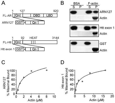
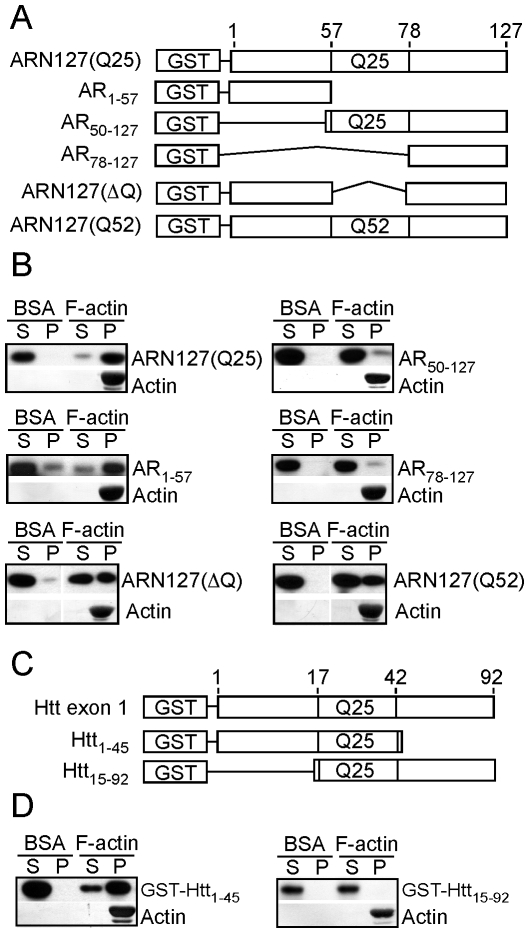
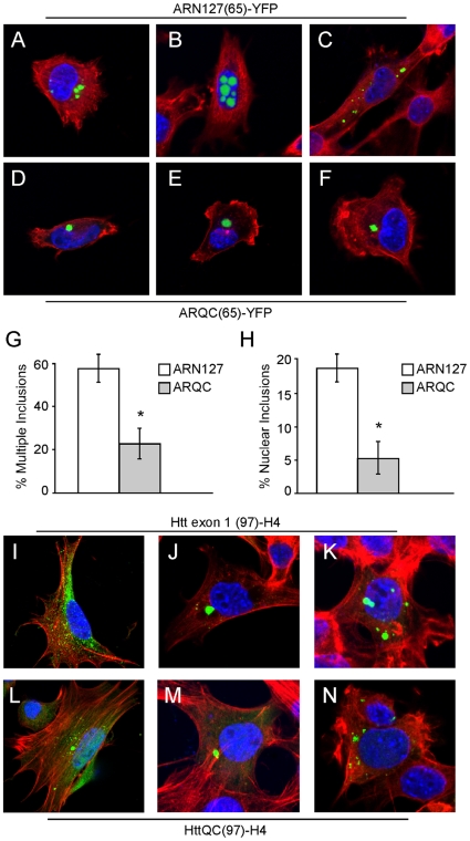
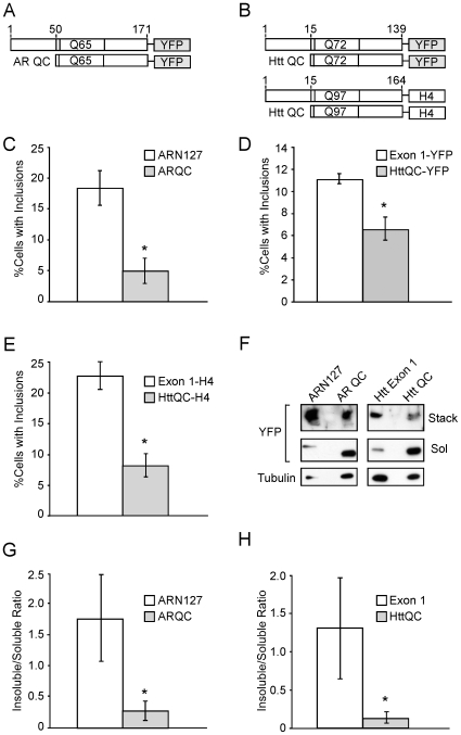
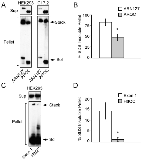
Protein aggregation is associated with neurodegeneration. Polyglutamine expansion diseases such as spinobulbar muscular atrophy and Huntington disease feature proteins that are destabilized by an expanded polyglutamine tract in their N-termini. It has previously been reported that intracellular aggregation of these target proteins, the androgen receptor (AR) and huntingtin (Htt), is modulated by actin-regulatory pathways. Sequences that flank the polyglutamine tract of AR and Htt might influence protein aggregation and toxicity through protein-protein interactions, but this has not been studied in detail. Here we have evaluated an N-terminal 127 amino acid fragment of AR and Htt exon 1. The first 50 amino acids of ARN127 and the first 14 amino acids of Htt exon 1 mediate binding to filamentous actin in vitro. Deletion of these actin-binding regions renders the polyglutamine-expanded forms of ARN127 and Htt exon 1 less aggregation-prone, and increases the SDS-solubility of aggregates that do form. These regions thus appear to alter the aggregation frequency and type of polyglutamine-induced aggregation. These findings highlight the importance of flanking sequences in determining the propensity of unstable proteins to misfold.


Conflict of interest statement

Competing Interests: The authors have declared that no competing interests exist.

Figures

Figure 1
Figure 1. ARN127 and Htt exon 1 directly bind to F-actin in vitro.
A, Schematic of GST-tagged N-terminal fragments of AR and Htt, GST-ARN127 and GST-Htt exon 1, in comparison to full-length AR (FL-AR) and full-length Htt (FL-Htt). B, GST-ARN127(25) and GST-Htt exon 1(25) co-sediment with F-actin in vitro but remain soluble in the absence of F-actin. 0.5 µM of precleared GST-ARN127(25) or .25 µM of precleared GST-Htt exon 1(25) was mixed with 4 µM of pre-polymerized F-actin. Mixtures were ultracentrifuged at 100,000 x g and supernatant and pellet fractions were analyzed via western blot (ARN127 and Htt exon 1) and Coomassie (actin). GST alone does not co-sediment with F-actin. C, Binding affinity of GST-ARN127(25) to F-actin. The F-actin co-sedimentation assay was performed with 0, .5 µM, 1 µM, 2 µM, 4 µM, and 8 µM F-actin and .016 µM GST-ARN127(25). Gels were quantified to determine the percent bound to F-actin. Percent maximal binding is reported. D, Binding affinity of GST-Htt exon 1(25) to F-actin. The F-actin co-sedimentation assay was performed with 0, .5 µM, 1 µM, 2 µM, 4 µM, 8 µM, and 16 µM F-actin and .15 µM GST-Htt exon 1(25). Gels were quantified to determine the percent bound to F-actin using Image J.
Figure 2
Figure 2. N50 of ARN127 and N14 of Htt exon 1 mediate F-actin binding in vitro.
A, Schematic of GST-ARN127 and various truncation mutants. B, The N-terminus of ARN127 binds to F-actin in vitro. GST-tagged truncations of ARN127(25) (AR1–57, AR50–127, AR78–127 (20 nM)), were tested for binding to F-actin (4 µM). ARN127(25) and AR1–57 co-sediment with F-actin while AR50–127 and AR78–127 do not. GST-tagged ARN127(ΔQ) or ARN127(52) (0.5 µM) both co-sediment with F-actin. C, Schematic of GST-Htt exon 1 and GST-tagged truncation mutants. D, The N-terminus of Htt exon 1 binds to F-actin in vitro. GST-tagged Htt1–45 (0.1 µM) binds to F-actin (4 µM) while Htt15–92 (0.1 µM) does not.
Figure 3
Figure 3. N50 of ARN127 mediates aggregation inhibition by actin- regulatory pathways.
A, Y-27632 inhibits aggregation of ARN127(65)-CFP/YFP and increases ARQC(65)-CFP/YFP aggregation. Cells expressing either ARN127 or ARQC fused to CFP/YFP were treated with 10, 30, or 100 µM Y-27632 for 24 hours. Relative FRET/donor measurements represent the change in aggregation from compounds compared to untreated cells. Y-27632 suppressed ARN127(65)-CFP/YFP aggregation, whereas it increased ARQC(65)-CFP/YFP aggregation. B, Profilin 1 decreases aggregation of ARN127(65)-CFP/YFP dose-dependently. Profilin 1 was cotransfected with polyglutamine proteins at increasing amounts, .075, .15, or .225 µg. This response was diminished for ARQC(65)-CFP/YFP. (* = p<.01, ** = p<.0001, Student's t-test).
Figure 4
Figure 4. N50 of ARN127 influences inclusion type, number, and distribution.
A–C, Confocal images of ARN127(65)-YFP inclusions in C17.2 cells at 48 h (60X). D–F, Confocal images of ARQC(65)-YFP inclusions in C17.2 cells at 48 h (60X). YFP-tagged proteins are in green, F-actin is stained with rhodamine-phalloidin (red) and DNA is stained with DAPI (blue). G, ARN127(65)-YFP more often forms multiple inclusions per cell than ARQC(65)-YFP (* = p<.002, Student's t-test). H, ARN127(Q65)-YFP more often forms nuclear inclusions per cell than ARQC(65)-YFP (* = p<.002, Student's t-test). Averages are from three separate transfections, counting at least 100 cells each. I-K, Confocal images of Htt exon 1(97)-H4 in C17.2 cells at 48 h (60X). L-N, Confocal images of HttQC(97)-H4 in C17.2 cells at 48 h (60X). Immunofluorescence of HA-tagged Htt is in green, F-actin is stained with rhodamine-phalloidin (red) and DNA is stained with DAPI (blue). We did not observe significant differences in patterns of inclusion formation between the two Htt constructs.
Figure 5
Figure 5. N50 of ARN127 and N14 of Htt exon 1 increase inclusion formation and detergent insolubility of aggregates.
HEK293 cells were transfected with the indicated constructs and evaluated after 24 h. A, Schematic of AR fusion proteins, not to scale. B, Schematic of Htt fusion proteins, not to scale. The H4 sequence represents HIS-HA-HA-HIS epitopes. C, ARN127(65)-YFP forms more inclusions than ARQC(65)-YFP (* = p<.0025, Student's t-test). D, Htt exon 1(72)-YFP forms more inclusions than HttQC(72)-YFP (* = p<.0025, Student's t-test). E, Htt exon 1(97)-H4 forms more inclusions than HttQC(97)-H4 (* = p<.005, Student's t-test). F, ARN127(65)-YFP and Htt exon 1(72)-YFP form more SDS-insoluble aggregates than ARQC(65)-YFP and HttQC(72)-YFP. HEK293 cells were transiently transfected with the indicated constructs. After 24 h, cells were lysed in 2% SDS sample buffer, and subjected to SDS-PAGE and western blot with YFP antibody. Stack indicates the SDS-insoluble higher molecular weight aggregates trapped in the stacking gel; Sol indicates the SDS-soluble monomers. Tubulin indicates loading control. Deletion of amino terminal peptides reduced the overall proportion of SDS-insoluble material detected in the stacking gel. G, Quantification of relative insoluble to soluble fractions of ARN127 vs. ARQC (n = 3, * = p<.05, Student's t-test). H, Quantification of relative insoluble to soluble fractions of Htt exon 1 vs. HttQC (n = 3, * = p<.05, Student's t-test). Quantification by Image J.
Figure 6
Figure 6. N50 of ARN127 and N14 of Htt exon 1 increase SDS-insoluble aggregate formation.
A, ARN127(65)-YFP forms more SDS-insoluble aggregates than ARQC(65)-YFP. HEK293 and C17.2 cells were transiently transfected with either ARN127(65)-YFP or ARQC(65)-YFP. After 24 h, cells were syringe lysed and fractionated by centrifugation at 15,000 x g. Supernatant and pellet fractions were then resuspended in 2% SDS sample buffer and subjected to SDS-PAGE and western blot with YFP antibody. The soluble portions are shown in the upper panels, while the insoluble portions are shown in the lower panels. Stack indicates the SDS-insoluble higher molecular weight aggregates in the stacking gel; Sol indicates the SDS-soluble monomers. B, Quantification of SDS-insoluble aggregates for ARN127(65)-YFP and ARQC(65)-YFP from HEK293 cells after 24 h. The insoluble fraction of ARN127(65)-YFP has significantly more SDS-insoluble material than ARQC(65)-YFP (n = 3, * = p<.001, Student's t-test). C, Htt exon1(97)-H4 forms more insoluble aggregates than HttQC(97)-H4 in HEK293 cells after 24 h. Cells were treated as above and subjected to SDS-PAGE and western blot with HA antibody. D, Quantification of SDS-insoluble aggregates for Htt exon1(97)-H4 and HttQC(97)-H4 from HEK293 cells after 24 h. The insoluble fraction of Htt exon 1(97)-H4 has significantly more SDS-insoluble material than HttQC(97)-H4 (n = 3, * = p<.005, Student's t-test).

Similar articles

Cited by

References

    1. La Spada AR, Wilson EM, Lubahn DB, Harding AE, Fischbeck KH. Androgen receptor gene mutations in X-linked spinal and bulbar muscular atrophy. Nature. 1991;352:77–79. - PubMed
    1. A novel gene containing a trinucleotide repeat that is expanded and unstable on Huntington's disease chromosomes. The Huntington's Disease Collaborative Research Group. Cell. 1993;72:971–983. - PubMed
    1. Li M, Chevalier-Larsen ES, Merry DE, Diamond MI. Soluble androgen receptor oligomers underlie pathology in a mouse model of spinobulbar muscular atrophy. J Biol Chem. 2007;282:3157–3164. - PubMed
    1. Goldberg YP, Nicholson DW, Rasper DM, Kalchman MA, Koide HB, et al. Cleavage of huntingtin by apopain, a proapoptotic cysteine protease, is modulated by the polyglutamine tract. Nat Genet. 1996;13:442–449. - PubMed
    1. Li M, Miwa S, Kobayashi Y, Merry DE, Yamamoto M, et al. Nuclear inclusions of the androgen receptor protein in spinal and bulbar muscular atrophy. Ann Neurol. 1998;44:249–254. - PubMed

Publication types

MeSH terms