. 2010 Feb 5;6(2):e1000835.
doi: 10.1371/journal.pgen.1000835.

DNA binding of centromere protein C (CENPC) is stabilized by single-stranded RNA

Affiliations

DNA binding of centromere protein C (CENPC) is stabilized by single-stranded RNA

Yaqing Du et al. PLoS Genet. .

Abstract

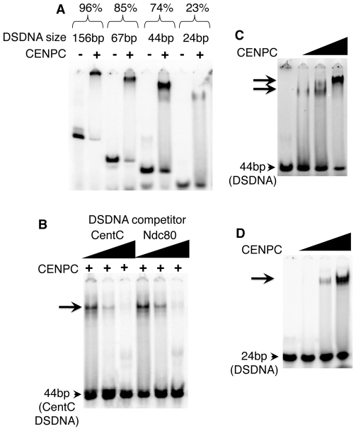
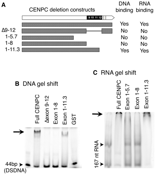
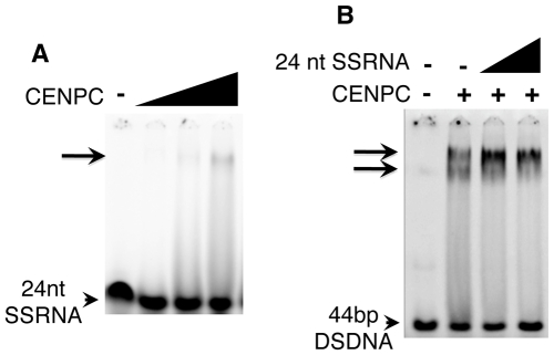
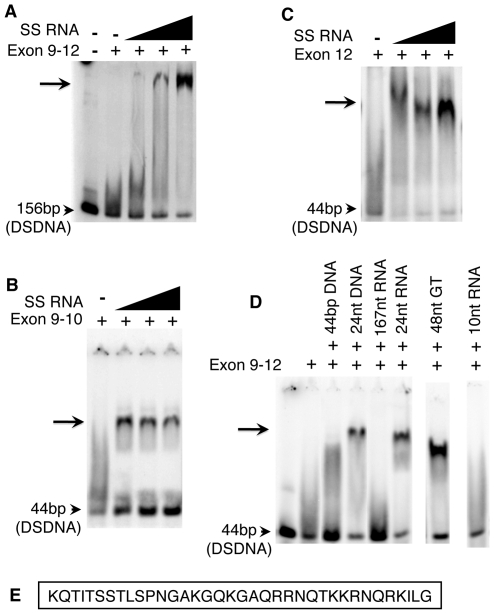
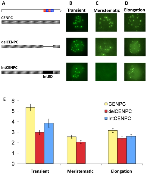
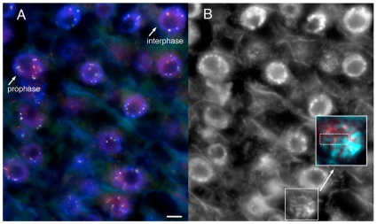
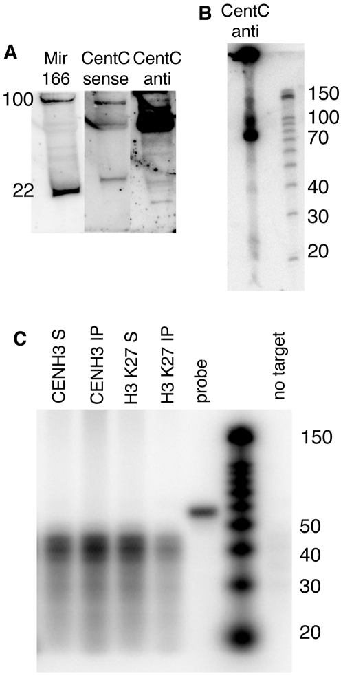
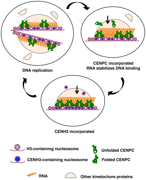
Centromeres are the attachment points between the genome and the cytoskeleton: centromeres bind to kinetochores, which in turn bind to spindles and move chromosomes. Paradoxically, the DNA sequence of centromeres has little or no role in perpetuating kinetochores. As such they are striking examples of genetic information being transmitted in a manner that is independent of DNA sequence (epigenetically). It has been found that RNA transcribed from centromeres remains bound within the kinetochore region, and this local population of RNA is thought to be part of the epigenetic marking system. Here we carried out a genetic and biochemical study of maize CENPC, a key inner kinetochore protein. We show that DNA binding is conferred by a localized region 122 amino acids long, and that the DNA-binding reaction is exquisitely sensitive to single-stranded RNA. Long, single-stranded nucleic acids strongly promote the binding of CENPC to DNA, and the types of RNAs that stabilize DNA binding match in size and character the RNAs present on kinetochores in vivo. Removal or replacement of the binding module with HIV integrase binding domain causes a partial delocalization of CENPC in vivo. The data suggest that centromeric RNA helps to recruit CENPC to the inner kinetochore by altering its DNA binding characteristics.


Conflict of interest statement

The authors have declared that no competing interests exist.

Figures

Figure 1
Figure 1. Maize CENPC binds to DNA.
(A) Purified full CENPC and different sized CentC DSDNA binding substrates (indicated at top). The binding efficiency as % binding is shown (bound DNA/total). (B) Competition experiments. Lanes were loaded with the mixture of full CENPC protein, 44 bp CentC DSDNA, and either unlabeled 44 bp CentC or 44 bp Ndc80 DSDNA. (C) Increasing quantities of CENPC cause a supershift on a 44 bp substrate. (D) Increasing quantities of CENPC do not cause a supershift on the 24 bp substrate. The triangles represent the amount of protein or DNA added, and arrows indicate the shifted bands.
Figure 2
Figure 2. DNA and RNA binding localize to exon 9–12.
(A) Schematic representation of CENPC constructs used. (B) DNA binding. Radiolabeled 44 bp DSDNA was incubated with equal amounts of the different CENPC fragments. A shifted band (arrow) was seen for full CENPC and Exon 1–11.3, but not for Δ Exon 9–12, Exon 1–5.7 or Exon 1–8. As a negative control, GST (Glutathione S-transferase) was tested and no band shift was seen. (C) RNA binding. Radiolabeled 167 nt RNA was incubated with equal amounts of different CENPC fragments. Free RNA appears as two bands as expected for a long RNA with double stranded character (arrowheads). A shifted band, which is too large to enter the gel matrix (but visible, see arrow), was seen for full CENPC and Exon 1–11.3, but not for Exon 1–5.7 or Exon 1–8.
Figure 3
Figure 3. SSRNA causes a supershift of the CENPC/DNA complex.
(A) 24 nt SSRNA binds weakly to CENPC. A faint shifted band is seen when CENPC is added at high concentrations. (B) Increasing amounts (triangles) of unlabeled 24 nt SSRNA cause the formation of a supershifted band similar to what is observed when excess CENPC protein is added (Figure 1C).
Figure 4
Figure 4. The purified Exon 9–12 domain requires RNA to bind DNA in vitro.
(A) The DNA binding domain of CENPC alone does not bind to DNA, however a shifted band becomes evident as increasing amounts of SSRNA are added. The same is true for Exon 9–10 (B) and exon 12 alone (C). (D) Only single-stranded RNA or DNA stabilizes Exon 9–12 for DSDNA binding. Gel shift is observed in the presence of 24 nt SSDNA, 24 nt SSRNA, and 48 nt poly-GT (SSDNA). However, no shift is observed for 44 bp DSDNA, 167 nt RNA (with double stranded character) or 10 nt RNA. (E) Sequence of CENPC exon 12.
Figure 5
Figure 5. Removal or replacement of Exon 9–12 delocalizes CENPC in vivo.
(A) Schematic representation of the YFP–CENPC constructs used in maize transformation. (B) Transient expression of YFP–CENPC in cultured cells. The green YFP spots represent kinetochore localization, as determined from fixed cells (Figure 6). (C) Fluorescence in root tips of stably transformed plants. (D) Fluorescence in the elongation zone of stably transformed plants. Images are projections showing all YFP signal (green) from single nuclei. (E) Quantification of the data in (B–D). The Y axis represents the ratio of YFP signal in kinetochores to YFP signal in the nucleoplasm. There are significant differences between full CENPC and delCENPC and IntCENPC in all three types of tissue (P<0.05, bars show SEM).
Figure 6
Figure 6. YFP–CENPC localizes to kinetochores.
(A) A cryostat section from a root tip, showing cells in various stages of the cell cycle. YFP–CENPC is labeled by anti–YFP antisera (red), while YFP itself is shown in green. The red and green signals overlay to produce a yellow color. DNA (DAPI) is shown in blue. Cells in interphase and prophase are noted and differentiated by chromatin staining. Kinetochores on prophase chromosomes are noted with arrows to show the paired spots on replicated chromatids. (B) A black and white version of the DNA stain in (A). An early prophase cell is enlarged in the panel to highlight a single chromosome, with anti–YFP staining (red) lying in the primary constriction. Each kinetochore is noted with an arrow. Bar = 5 µm.
Figure 7
Figure 7. Chromatin-associated CentC transcripts are predominantly 75 nt and do not include siRNAs.
(A) Northern blot of total maize RNA. DNase-treated total RNA (enriched for ≤200 bp) was separated by PAGE and blotted to a membrane. Radiolabeled RNA probes specific to microRNA166 (miR166) and CentC forward (GenBank AY530283.1) and reverse strands were hybridized in succession. The mature form of miR166 is 22 nt while its precursors are ∼100 nt and seen near the top of the gel. Molecular weights were estimated by ethidium bromide staining of 22 and 28 nt RNA oligonucleotides (not shown). (B) Higher resolution RNA blot showing the size classes of CentC forward transcripts. Molecular markers are shown on the right. (C) Analysis of RNA associated with CENH3 and H3-containing chromatin. RNA recovered from ChIP experiments (3.34 µg) was subjected to RNase protection (RPA) using the CentC forward probe (52 nt). The upper resolution of detectable sizes is 44 nt (8 nt less than the probe size). The fraction associated with immune complex is labeled ‘IP’ and the non-associated fraction is labeled ‘S’. Yeast RNA was used to demonstrate complete digestion in the absence of target DNA, and is labeled ‘no target’.
Figure 8
Figure 8. A model for how RNA facilitates the CENPC-DNA interaction.
Three stages of kinetochore replication are shown in a cycle that broadly represents the cell cycle. DNA replication splits kinetochores and distributes the resident kinetochore proteins to sister chromatids. CENPC recruitment is continuous. Protein–protein contacts bring CENPC to the kinetochore while resident RNA stabilizes CENPC for DNA binding. CENH3 assembly is a discrete event that occurs after S phase, as early as G2 or as late as anaphase or G1 ,. CENPC and CENH3 assembly are separated in time such that one can guide the other.

Similar articles

Cited by

  • The Genomic Landscape of Centromeres in Cancers.
    Saha AK, Mourad M, Kaplan MH, Chefetz I, Malek SN, Buckanovich R, Markovitz DM, Contreras-Galindo R. Saha AK, et al. Sci Rep. 2019 Aug 2;9(1):11259. doi: 10.1038/s41598-019-47757-6. Sci Rep. 2019. PMID: 31375789 Free PMC article.
  • Epigenetic regulation of centromere function.
    Wong CYY, Lee BCH, Yuen KWY. Wong CYY, et al. Cell Mol Life Sci. 2020 Aug;77(15):2899-2917. doi: 10.1007/s00018-020-03460-8. Epub 2020 Feb 1. Cell Mol Life Sci. 2020. PMID: 32008088 Free PMC article. Review.
  • Repetitive centromeric satellite RNA is essential for kinetochore formation and cell division.
    Rošić S, Köhler F, Erhardt S. Rošić S, et al. J Cell Biol. 2014 Nov 10;207(3):335-49. doi: 10.1083/jcb.201404097. Epub 2014 Nov 3. J Cell Biol. 2014. PMID: 25365994 Free PMC article.
  • Genome-wide mapping reveals R-loops associated with centromeric repeats in maize.
    Liu Y, Liu Q, Su H, Liu K, Xiao X, Li W, Sun Q, Birchler JA, Han F. Liu Y, et al. Genome Res. 2021 Aug;31(8):1409-1418. doi: 10.1101/gr.275270.121. Epub 2021 Jul 9. Genome Res. 2021. PMID: 34244230 Free PMC article.
  • Genetic drivers and cellular selection of female mosaic X chromosome loss.
    Liu A, Genovese G, Zhao Y, Pirinen M, Zekavat SM, Kentistou KA, Yang Z, Yu K, Vlasschaert C, Liu X, Brown DW, Hudjashov G, Gorman BR, Dennis J, Zhou W, Momozawa Y, Pyarajan S, Tuzov V, Pajuste FD, Aavikko M, Sipilä TP, Ghazal A, Huang WY, Freedman ND, Song L, Gardner EJ; FinnGen; Estonian Biobank Research Team; Breast Cancer Association Consortium; Million Veteran Program; Sankaran VG, Palotie A, Ollila HM, Tukiainen T, Chanock SJ, Mägi R, Natarajan P, Daly MJ, Bick A, McCarroll SA, Terao C, Loh PR, Ganna A, Perry JRB, Machiela MJ. Liu A, et al. Nature. 2024 Jul;631(8019):134-141. doi: 10.1038/s41586-024-07533-7. Epub 2024 Jun 12. Nature. 2024. PMID: 38867047

References

    1. Yu HG, Hiatt EN, Dawe RK. The plant kinetochore. Trends Plant Sci. 2000;5:543–547. - PubMed
    1. Kline-Smith SL, Sandall S, Desai A. Kinetochore-spindle microtubule interactions during mitosis. Curr Opin Cell Biol. 2005;17:35–46. - PMC - PubMed
    1. Kotwaliwale C, Biggins S. Microtubule capture: a concerted effort. Cell. 2006;127:1105–1108. - PubMed
    1. Fukagawa T. The kinetochore and spindle checkpoint in vertebrate cells. Front Biosci. 2008;13:2705–2713. - PubMed
    1. Henikoff S, Ahmad K, Malik HS. The centromere paradox: stable inheritance with rapidly evolving DNA. Science. 2001;293:1098–1102. - PubMed

Publication types

MeSH terms