

Mutations in SLC29A3, encoding an equilibrative nucleoside transporter ENT3, cause a familial histiocytosis syndrome (Faisalabad histiocytosis) and familial Rosai-Dorfman disease
- PMID: 20140240
- PMCID: PMC2816679
- DOI: 10.1371/journal.pgen.1000833
Mutations in SLC29A3, encoding an equilibrative nucleoside transporter ENT3, cause a familial histiocytosis syndrome (Faisalabad histiocytosis) and familial Rosai-Dorfman disease
Abstract
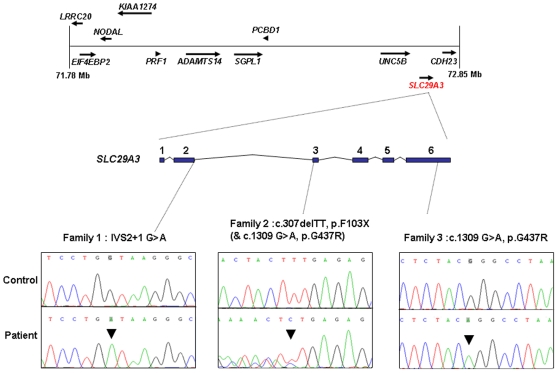
The histiocytoses are a heterogeneous group of disorders characterised by an excessive number of histiocytes. In most cases the pathophysiology is unclear and treatment is nonspecific. Faisalabad histiocytosis (FHC) (MIM 602782) has been classed as an autosomal recessively inherited form of histiocytosis with similarities to Rosai-Dorfman disease (RDD) (also known as sinus histiocytosis with massive lymphadenopathy (SHML)). To elucidate the molecular basis of FHC, we performed autozygosity mapping studies in a large consanguineous family and identified a novel locus at chromosome 10q22.1. Mutation analysis of candidate genes within the target interval identified biallelic germline mutations in SLC29A3 in the FHC kindred and in two families reported to have familial RDD. Analysis of SLC29A3 expression during mouse embryogenesis revealed widespread expression by e14.5 with prominent expression in the central nervous system, eye, inner ear, and epithelial tissues including the gastrointestinal tract. SLC29A3 encodes an intracellular equilibrative nucleoside transporter (hENT3) with affinity for adenosine. Recently germline mutations in SLC29A3 were also described in two rare autosomal recessive disorders with overlapping phenotypes: (a) H syndrome (MIM 612391) that is characterised by cutaneous hyperpigmentation and hypertrichosis, hepatomegaly, heart anomalies, hearing loss, and hypogonadism; and (b) PHID (pigmented hypertrichosis with insulin-dependent diabetes mellitus) syndrome. Our findings suggest that a variety of clinical diagnoses (H and PHID syndromes, FHC, and familial RDD) can be included in a new diagnostic category of SLC29A3 spectrum disorder.
Conflict of interest statement
The authors have declared that no competing interests exist.
Figures


Similar articles
-
Phenotypic intrafamilial variability including H syndrome and Rosai-Dorfman disease associated with the same c.1088G > A mutation in the SLC29A3 gene.Hum Genomics. 2021 Oct 17;15(1):63. doi: 10.1186/s40246-021-00362-z. Hum Genomics. 2021. PMID: 34657628 Free PMC article.
-
A case of H syndrome showing immunophenotye similarities to Rosai-Dorfman disease.Am J Dermatopathol. 2011 Feb;33(1):47-51. doi: 10.1097/DAD.0b013e3181ee547c. Am J Dermatopathol. 2011. PMID: 21178579
-
An Egyptian family with H syndrome due to a novel mutation in SLC29A3 illustrating overlapping features with pigmented hypertrichotic dermatosis with insulin-dependent diabetes and Faisalabad histiocytosis.Pediatr Diabetes. 2013 Sep;14(6):466-72. doi: 10.1111/j.1399-5448.2012.00925.x. Epub 2012 Sep 18. Pediatr Diabetes. 2013. PMID: 22989030
-
Equilibrative nucleotide transporter ENT3 (SLC29A3): A unique transporter for inherited disorders and cancers.Exp Cell Res. 2024 Jan 15;434(2):113892. doi: 10.1016/j.yexcr.2023.113892. Epub 2023 Dec 16. Exp Cell Res. 2024. PMID: 38104646 Review.
-
A novel start-loss mutation of the SLC29A3 gene in a consanguineous family with H syndrome: clinical characteristics, in silico analysis and literature review.BMC Med Genomics. 2024 Jul 4;17(1):178. doi: 10.1186/s12920-024-01949-w. BMC Med Genomics. 2024. PMID: 38965556 Free PMC article. Review.
Cited by
-
A mild form of SLC29A3 disorder: a frameshift deletion leads to the paradoxical translation of an otherwise noncoding mRNA splice variant.PLoS One. 2012;7(1):e29708. doi: 10.1371/journal.pone.0029708. Epub 2012 Jan 4. PLoS One. 2012. PMID: 22238637 Free PMC article.
-
Diagnosis and treatment of Rosai-Dorfman disease of the spine: a systematic literature review.Syst Rev. 2021 Jan 18;10(1):31. doi: 10.1186/s13643-021-01581-0. Syst Rev. 2021. PMID: 33461611 Free PMC article.
-
Update on the Genetics of Autoinflammatory Disorders.Curr Allergy Asthma Rep. 2019 Jul 18;19(9):41. doi: 10.1007/s11882-019-0874-2. Curr Allergy Asthma Rep. 2019. PMID: 31321571 Review.
-
Rosai-Dorfman disease as a rare cause of cervical lymphadenopathy - case report and literature review.Cent Eur J Immunol. 2018;43(3):341-345. doi: 10.5114/ceji.2018.80055. Epub 2018 Oct 30. Cent Eur J Immunol. 2018. PMID: 30588179 Free PMC article. Review.
-
Purine import into malaria parasites as a target for antimalarial drug development.Ann N Y Acad Sci. 2015 Apr;1342(1):19-28. doi: 10.1111/nyas.12568. Epub 2014 Nov 25. Ann N Y Acad Sci. 2015. PMID: 25424653 Free PMC article. Review.
References
-
- Cline MJ. Histiocytes and histiocytosis. Blood. 1994;84(9):2840–2853. - PubMed
-
- Writing group of the Histiocyte society. Histiocytosis syndromes in children. Lancet. 1987;1:208–209. - PubMed
-
- Zur Stadt U, Beutel K, Kolberg S, Schneppenheim R, Kabisch H, et al. Mutation spectrum in children with primary hemophagocytic lymphohistiocytosis: molecular and functional analyses of PRF1, UNC13D, STX11, and RAB27A. Hum Mut. 2006;27(1):62–68. - PubMed
Publication types
MeSH terms
Substances
Grants and funding
LinkOut - more resources
Full Text Sources
Medical
Molecular Biology Databases
