. 2010 Feb 3;5(2):e9006.
doi: 10.1371/journal.pone.0009006.

Structure and kinetic investigation of Streptococcus pyogenes family GH38 alpha-mannosidase

Affiliations

Structure and kinetic investigation of Streptococcus pyogenes family GH38 alpha-mannosidase

Michael D L Suits et al. PLoS One. .

Abstract

Background: The enzymatic hydrolysis of alpha-mannosides is catalyzed by glycoside hydrolases (GH), termed alpha-mannosidases. These enzymes are found in different GH sequence-based families. Considerable research has probed the role of higher eukaryotic "GH38" alpha-mannosides that play a key role in the modification and diversification of hybrid N-glycans; processes with strong cellular links to cancer and autoimmune disease. The most extensively studied of these enzymes is the Drosophila GH38 alpha-mannosidase II, which has been shown to be a retaining alpha-mannosidase that targets both alpha-1,3 and alpha-1,6 mannosyl linkages, an activity that enables the enzyme to process GlcNAc(Man)(5)(GlcNAc)(2) hybrid N-glycans to GlcNAc(Man)(3)(GlcNAc)(2). Far less well understood is the observation that many bacterial species, predominantly but not exclusively pathogens and symbionts, also possess putative GH38 alpha-mannosidases whose activity and specificity is unknown.

Methodology/principal findings: Here we show that the Streptococcus pyogenes (M1 GAS SF370) GH38 enzyme (Spy1604; hereafter SpGH38) is an alpha-mannosidase with specificity for alpha-1,3 mannosidic linkages. The 3D X-ray structure of SpGH38, obtained in native form at 1.9 A resolution and in complex with the inhibitor swainsonine (K(i) 18 microM) at 2.6 A, reveals a canonical GH38 five-domain structure in which the catalytic "-1" subsite shows high similarity with the Drosophila enzyme, including the catalytic Zn(2+) ion. In contrast, the "leaving group" subsites of SpGH38 display considerable differences to the higher eukaryotic GH38s; features that contribute to their apparent specificity.

Conclusions/significance: Although the in vivo function of this streptococcal GH38 alpha-mannosidase remains unknown, it is shown to be an alpha-mannosidase active on N-glycans. SpGH38 lies on an operon that also contains the GH84 hexosaminidase (Spy1600) and an additional putative glycosidase. The activity of SpGH38, together with its genomic context, strongly hints at a function in the degradation of host N- or possibly O-glycans. The absence of any classical signal peptide further suggests that SpGH38 may be intracellular, perhaps functioning in the subsequent degradation of extracellular host glycans following their initial digestion by secreted glycosidases.


Conflict of interest statement

Competing Interests: The authors have declared that no competing interests exist.

Figures

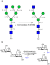
Figure 1
Figure 1. Catalytic activity of GH38 α−mannosidases.
(A) Golgi α−mannosidase II is responsible for the hydrolysis of both α−1,3 and α−1–6 mannosides during the diversification of hybrid N glycans (GlcNAcMan5GlcNAc2 becoming GlcNAcMan3GlcNAc2). (B) The catalytic action of a retaining α−mannosidase, here exemplified for the α−1,3 mannosidase activity of GH38 enzymes; catalysis occurs with net retention of anomeric configuration.
Figure 2
Figure 2. Catalytic activity of SpGH38 α−mannosidase and inhibition by swainsonine.
(A) Activity on 4-methylumbelliferyl α−D mannoside (4-MeUMB) and (B) α−1,3 mannobiose (see text for details) Substrate insolubility under the conditions used precluded higher [S] values. (C) The K i for swainsonine was determined using α−1,3 mannobiose as substrate with [S] < < Km and [I] straddling the K i. V0 and Vi are the rates of the reaction in the absence and presence of inhibitor, respectively. The K i for a competitive inhibitor is derived from the gradient of 1/K i (see Methods ); here 18±0.5 µM.
Figure 3
Figure 3. SpGH38-catalysed hydrolysis of Man9(GlcNAc)2 glycans.
(A) Action of SpGH38, alone, on Man9(GlcNAc)2. The glycan remains unmodified. (B) Action of SpGH38 in combination with a specific α−1,2 mannosidase the Bacteroides thetaiotaomicron Bt3990. Following α−1,2 mannoside removal (which has previously been shown to be specific, see Supplemental Figure 1 in , SpGH38 is able to further degrade the unmasked glycans, with the action pattern most indicative of α−1,3 mannosidase activity. An α−1,3 mannosidase activity for SpGH38 is further supported by the specificity of the enzyme for the disaccharide α−1,3 mannobiose (see text).
Figure 4
Figure 4. 3-D structure of SpGH38 and its swainsonine complex.
(A) 3-D topology cartoon (divergent stereo) colored according to domains with swainsonine in ball-and-stick. N-term (red: 1–294) 3-α (green: 295–392), β-1 (blue: 393–514,806–825), β−2 (yellow: 522–805) and β−3 (cyan: 825–901) (B) Surface representation of SpGH38 colored as for part (A). (C) Active centre and electron density for the Swainsonine/Zn2+ complex of SpGH38 (divergent stereo). The map shown is the unbiased Fobs-Fcalc synthesis, contoured at 2.5 σ, calculated with model phases prior to the incorporation of Swainsonine/Zn2+ in any refinement. (D) schematic diagram of the interactions of swainsonine (shown in panel C) with H-bonds >3.0 Å shown as dashed lines and residue numbers for the SpGH38 indicated. Arg149 makes a close contact to a swainsonine carbon (indicated with an arrow) of 2.9 Å (spatially equivalent to an H-bond to mannose O6 of the true substrate).
Figure 5
Figure 5. Conservation of GH38 reaction mechanism.
(A) Conserved active-centre constellation (here -1 subsite only) between the SpGH38 (grey), bovine bLAM (cyan) and the Drosophila GH38 α−mannosidase (blue). (B). GH38 α−mannosidases are known to act with net retention of anomeric configuration; a mechanism in which a glycosyl-enzyme intermediate is flanked by oxocarbenium-ion like transition-states. The intermediate has been trapped by the Withers and Rose groups and shown to bind in a 1S5 skew-boat conformation which in the absence of evidence to the contrary might imply a transition-state close to a B2,5. Pseudo-Michaelis complexes published on the Drosophila α−mannosidase II show the −1 sugar in a 4C1 chair conformation but these have been obtained on a nucleophile-alanine variant so their conformational relevance to catalysis is unclear .
Figure 6
Figure 6. Substrate specificity in SpGH38.
An overlap of the Drosophila GH38 α−mannosidase II complexes with α−1,3 (green bonds) and α−1,6 linked ligands (blue bonds) with the SpGH38 structure (grey surface) focussing on the +1 subsite (the -1 subsites are essentially identical, Fig. 5A). Features which may contribute to 1,3 specificity include the position of W764, the interactions afforded by D763 and the tightness of the “sphincter” formed by D763 and the catalytic acid/base D232. The figure is shown in divergent (“wall-eyed”) stereo.

Similar articles

Cited by

References

    1. Davis BG. Hand in Glove - Investigating Glycocode. Chem Ind. 2000:134–138.
    1. Ito Y, Ogawa T. A novel approach to the stereoselective synthesis of beta-mannosides. Angew Chemie Int Ed. 1994;33:1765–1767.
    1. Crich D, Chandrasekera NS. Mechanism of 4,6-0-benzylidene-directed beta-Mannosylation as determined by alpha-deuterium kinetic isotope effects. Angew Chemie Int Ed. 2004;43:5386–5389. - PubMed
    1. Gridley JJ, Osborn HMI. Recent advances in the construction of beta-D-mannose and beta-D-mannosamine linkages. J Chem Soc Perkin Trans 1. 2000;10:1471–1491.
    1. Cantarel BL, Coutinho PM, Rancurel C, Bernard T, Lombard V, et al. The Carbohydrate-Active EnZymes database (CAZy): an expert resource for Glycogenomics. Nucleic Acids Res. 2009;37:D233–D238. - PMC - PubMed

Publication types

MeSH terms