. 2010 Feb 3;5(2):e9025.
doi: 10.1371/journal.pone.0009025.

Complete-proteome mapping of human influenza A adaptive mutations: implications for human transmissibility of zoonotic strains

Affiliations

Complete-proteome mapping of human influenza A adaptive mutations: implications for human transmissibility of zoonotic strains

Olivo Miotto et al. PLoS One. .

Abstract

Background: There is widespread concern that H5N1 avian influenza A viruses will emerge as a pandemic threat, if they become capable of human-to-human (H2H) transmission. Avian strains lack this capability, which suggests that it requires important adaptive mutations. We performed a large-scale comparative analysis of proteins from avian and human strains, to produce a catalogue of mutations associated with H2H transmissibility, and to detect their presence in avian isolates.

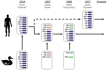
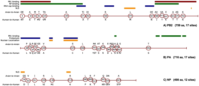
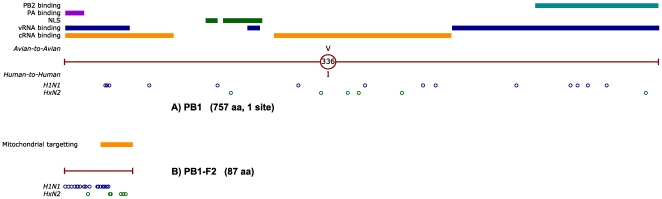
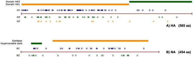
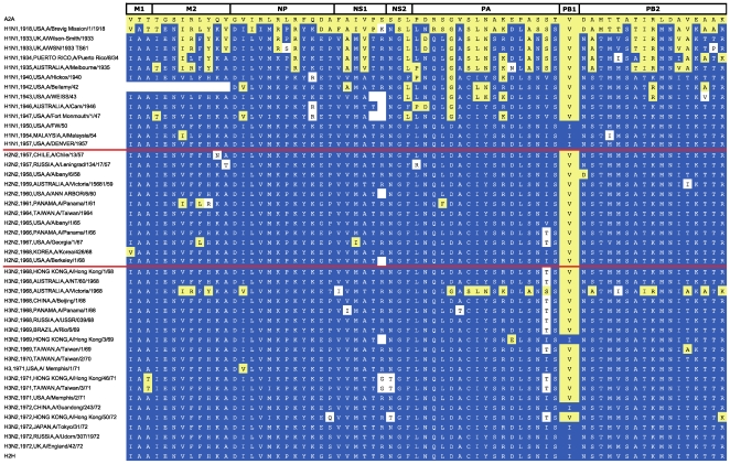
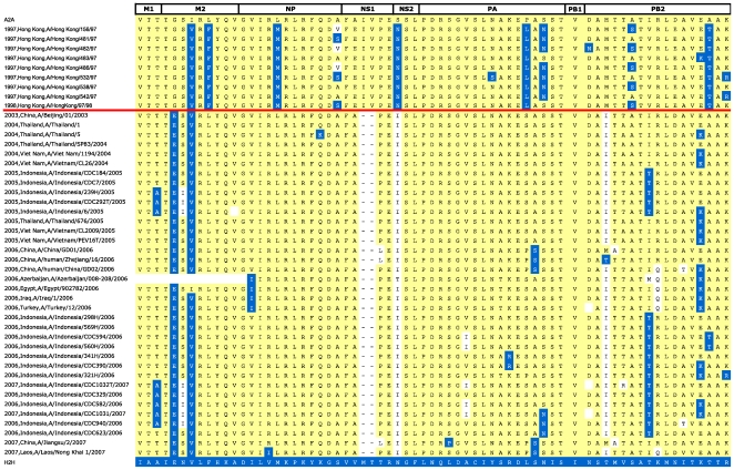
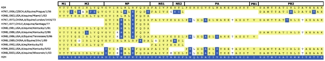
Methodology/principal findings: We constructed a dataset of influenza A protein sequences from 92,343 public database records. Human and avian sequence subsets were compared, using a method based on mutual information, to identify characteristic sites where human isolates present conserved mutations. The resulting catalogue comprises 68 characteristic sites in eight internal proteins. Subtype variability prevented the identification of adaptive mutations in the hemagglutinin and neuraminidase proteins. The high number of sites in the ribonucleoprotein complex suggests interdependence between mutations in multiple proteins. Characteristic sites are often clustered within known functional regions, suggesting their functional roles in cellular processes. By isolating and concatenating characteristic site residues, we defined adaptation signatures, which summarize the adaptive potential of specific isolates. Most adaptive mutations emerged within three decades after the 1918 pandemic, and have remained remarkably stable thereafter. Two lineages with stable internal protein constellations have circulated among humans without reassorting. On the contrary, H5N1 avian and swine viruses reassort frequently, causing both gains and losses of adaptive mutations.

Conclusions: Human host adaptation appears to be complex and systemic, involving nearly all influenza proteins. Adaptation signatures suggest that the ability of H5N1 strains to infect humans is related to the presence of an unusually high number of adaptive mutations. However, these mutations appear unstable, suggesting low pandemic potential of H5N1 in its current form. In addition, adaptation signatures indicate that pandemic H1N1/09 strain possesses multiple human-transmissibility mutations, though not an unusually high number with respect to swine strains that infected humans in the past. Adaptation signatures provide a novel tool for identifying zoonotic strains with the potential to infect humans.


Conflict of interest statement

Competing Interests: The authors have declared that no competing interests exist.

Figures

Figure 1
Figure 1. Human Influenza A reassortment events of the 20th Century.
The reassortment events associated with human pandemics in the 20th century (adapted from Webster et al. [39]). A full complement of eight gene segments of zoonotic origin caused the 1918 Spanish flu. In 1957, the H2N2 Asian flu pandemic replaced the HA, NA and PB1 segments, and in 1968 the H3N2 Hong Kong pandemic replaced the HA and PB1 segments only . In both cases, the new subtype fully replaced the subtypes previously circulating amongst humans. The 1977 Russian epidemic introduced an H1N1 strain almost identical to that circulating prior to 1957, and may have been caused by the release of 20-year old frozen viruses . The H1N1 and HxN2 lineages have since co-circulated in the human population; recently, their reassortment has given rise to human strains of H1N2 subtype.
Figure 2
Figure 2. Characteristic sites identified in components of the RNP assembly of influenza A (PB2, PA, NP proteins).
Circular markers, indicating the position of characteristic sites, are placed along the sequence length of the PB2 (A), PA (B) and NP (C) proteins of influenza A. Avian (A2A) variants are indicated above each marker, while human (H2H) variants are located below. If multiple characteristic variants are present, they are shown in decreasing order of frequency. In the upper part of each figure, colored lines show reported functional domains of PB2 –, PA – and NP .
Figure 3
Figure 3. Characteristic sites identified in the PB1 (A) and PB1-F2 (B) proteins of influenza A.
RNA segment 2, which encodes both the PB1 and PB1-F2 proteins, has been replaced at the onset of the 1957 and 1968 pandemics (see Figure 1). As a result, the H1N1 and HxN2 lineages do not share recent common origin for this segment. Characteristic mutations are therefore shown separately for the two lineages, in the lower part of each diagram, using blue (H1N1) and green (HxN2) circles. Known functional sites for PB1 , – and PB1-F2 are also indicated by colored lines in the upper part of each figure.
Figure 4
Figure 4. Characteristic sites identified in the matrix proteins M1 (A) and M2 (B) and non-structural proteins NS1 (C) and NEP/NS2 (D) of influenza A.
Identified characteristic sites are mapped against known functional domains of M1 , , M2 , NS1 – and NEP/NS2 , , using the notation used in Figure 2.
Figure 5
Figure 5. Characteristic sites identified in the HA (A) and NA (B) glycoproteins of influenza A.
The characteristic mutations identified for each of the subtypes present in humans are shown: H1 (blue circles), H2 (green circles), H3 (orange circles) for HA; and N1 (blue circles), N2 (green circles) for NA (details of these sites are given in Tables S1, S2, S3, S4, S5 of the Supplementary Materials S1). Known domains of these two proteins are indicated by coloured lines in the upper part of each figure.
Figure 6
Figure 6. Timeline of adaptation to H2H transmission for the influenza A proteome.
Adaptation signatures from human isolates between 1918 and 1972 are arranged in chronological order. Subtype, year and country of isolation, and isolate name are shown in the first column. The remaining columns show residues at all characteristic sites, in the order given in Table 4. A2A characteristic mutations are shown on a dark blue background, H2H mutations on a yellow background, while all other variants are on white. Blank cells represent unknown residues in incompletely sequenced proteomes. Consensus signatures for A2A and H2H proteomes are shown in the first and last row, respectively. Red horizontal lines indicate the start of the 1957 and 1968 pandemics, which introduced the H2N2 and H3N2 subtypes respectively.
Figure 7
Figure 7. Adaptation signatures of human-isolated H5N1 influenza A proteomes.
This figure shows the adaptation signatures of H5N1 sequences that infected humans in the period 1997–2008. For display clarity and conciseness, only a selection of representative signatures is presented. The same coloring scheme was used as in Figure 6. Dashes (in the NS1 protein signature) indicate amino acid deletions. A red horizontal line separates the early wave of infections in Hong Kong (1997–8) from more recent South-East Asian infections (since 2003).
Figure 8
Figure 8. Adaptation signatures of selected swine influenza A proteomes.
H2H signatures for a number of representative swine proteomes are shown. Subtype, year and country of isolation, and isolate name are shown in the first column, while the remaining columns show the signature residues, using the same coloring scheme as in Figure 6. The isolates are shown in four groups, according to signature similarity: (A) isolates with signatures similar to that of “classical swine” influenza, such as A/Swine/Iowa/15/30; (B) isolates from the H1N1/09 pandemic; (C) isolates with signatures that are very similar to those of contemporarily circulating human isolates; (D) isolates with predominantly “avian” signatures, containing very few or no H2H adaptive mutations. In most of groups, reassortment events are evidenced by discontinuities in the signature timeline.
Figure 9
Figure 9. Adaptation signatures of selected equine influenza A proteomes.
The signatures of selected equine isolates, spanning a period of nearly 50 years are shown. For conciseness, a number of similar signatures were removed from this set. Subtype, year and country of isolation, and isolate name are shown in the first column, while the remaining columns show the signature residues, using the same colouring scheme as in Figure 6.

Comment in

Similar articles

Cited by

References

    1. Taubenberger JK, Morens DM. Pandemic influenza–including a risk assessment of H5N1. Rev Sci Tech. 2009;28(1):187–202. - PMC - PubMed
    1. Potter CW. A history of influenza. J Appl Microbiol. 2001;91:572–579. - PubMed
    1. Capua I, Alexander DJ. Avian influenza and human health. Acta Trop. 2002;83(1):1–6. - PubMed
    1. Fraser C, Donnelly CA, Cauchemez S, Hanage WP, Van Kerkhove MD, et al. Pandemic potential of a strain of influenza A (H1N1): early findings. Science. 2009;324(5934):1557–1561. - PMC - PubMed
    1. Peiris JS, Tu WW, Yen HL. A novel H1N1 virus causes the first pandemic of the 21(st) Century. Eur J Immunol (Epub ahead of print, Sep 29) 2009 - PubMed

Publication types

MeSH terms