High-precision radiosurgical dose delivery by interlaced microbeam arrays of high-flux low-energy synchrotron X-rays
- PMID: 20140254
- PMCID: PMC2815784
- DOI: 10.1371/journal.pone.0009028
High-precision radiosurgical dose delivery by interlaced microbeam arrays of high-flux low-energy synchrotron X-rays
Abstract
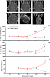
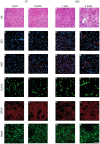
Microbeam Radiation Therapy (MRT) is a preclinical form of radiosurgery dedicated to brain tumor treatment. It uses micrometer-wide synchrotron-generated X-ray beams on the basis of spatial beam fractionation. Due to the radioresistance of normal brain vasculature to MRT, a continuous blood supply can be maintained which would in part explain the surprising tolerance of normal tissues to very high radiation doses (hundreds of Gy). Based on this well described normal tissue sparing effect of microplanar beams, we developed a new irradiation geometry which allows the delivery of a high uniform dose deposition at a given brain target whereas surrounding normal tissues are irradiated by well tolerated parallel microbeams only. Normal rat brains were exposed to 4 focally interlaced arrays of 10 microplanar beams (52 microm wide, spaced 200 microm on-center, 50 to 350 keV in energy range), targeted from 4 different ports, with a peak entrance dose of 200Gy each, to deliver an homogenous dose to a target volume of 7 mm(3) in the caudate nucleus. Magnetic resonance imaging follow-up of rats showed a highly localized increase in blood vessel permeability, starting 1 week after irradiation. Contrast agent diffusion was confined to the target volume and was still observed 1 month after irradiation, along with histopathological changes, including damaged blood vessels. No changes in vessel permeability were detected in the normal brain tissue surrounding the target. The interlacing radiation-induced reduction of spontaneous seizures of epileptic rats illustrated the potential pre-clinical applications of this new irradiation geometry. Finally, Monte Carlo simulations performed on a human-sized head phantom suggested that synchrotron photons can be used for human radiosurgical applications. Our data show that interlaced microbeam irradiation allows a high homogeneous dose deposition in a brain target and leads to a confined tissue necrosis while sparing surrounding tissues. The use of synchrotron-generated X-rays enables delivery of high doses for destruction of small focal regions in human brains, with sharper dose fall-offs than those described in any other conventional radiation therapy.
Conflict of interest statement
Figures


Similar articles
-
Effects of pulsed, spatially fractionated, microscopic synchrotron X-ray beams on normal and tumoral brain tissue.Mutat Res. 2010 Apr-Jun;704(1-3):160-6. doi: 10.1016/j.mrrev.2009.12.003. Epub 2009 Dec 23. Mutat Res. 2010. PMID: 20034592 Review.
-
Medical physics aspects of the synchrotron radiation therapies: Microbeam radiation therapy (MRT) and synchrotron stereotactic radiotherapy (SSRT).Phys Med. 2015 Sep;31(6):568-83. doi: 10.1016/j.ejmp.2015.04.016. Epub 2015 Jun 1. Phys Med. 2015. PMID: 26043881 Review.
-
Interlaced x-ray microplanar beams: a radiosurgery approach with clinical potential.Proc Natl Acad Sci U S A. 2006 Jun 20;103(25):9709-14. doi: 10.1073/pnas.0603567103. Epub 2006 Jun 7. Proc Natl Acad Sci U S A. 2006. PMID: 16760251 Free PMC article.
-
Response of avian embryonic brain to spatially segmented x-ray microbeams.Cell Mol Biol (Noisy-le-grand). 2001 May;47(3):485-93. Cell Mol Biol (Noisy-le-grand). 2001. PMID: 11441956
-
Permeability of Brain Tumor Vessels Induced by Uniform or Spatially Microfractionated Synchrotron Radiation Therapies.Int J Radiat Oncol Biol Phys. 2017 Aug 1;98(5):1174-1182. doi: 10.1016/j.ijrobp.2017.03.025. Epub 2017 Mar 21. Int J Radiat Oncol Biol Phys. 2017. PMID: 28721902
Cited by
-
Microbeam radiation therapy alters vascular architecture and tumor oxygenation and is enhanced by a galectin-1 targeted anti-angiogenic peptide.Radiat Res. 2012 Jun;177(6):804-12. doi: 10.1667/rr2784.1. Epub 2012 May 18. Radiat Res. 2012. PMID: 22607585 Free PMC article.
-
Non-conventional Ultra-High Dose Rate (FLASH) Microbeam Radiotherapy Provides Superior Normal Tissue Sparing in Rat Lung Compared to Non-conventional Ultra-High Dose Rate (FLASH) Radiotherapy.Cureus. 2021 Nov 6;13(11):e19317. doi: 10.7759/cureus.19317. eCollection 2021 Nov. Cureus. 2021. PMID: 35223216 Free PMC article.
-
Early gene expression analysis in 9L orthotopic tumor-bearing rats identifies immune modulation in molecular response to synchrotron microbeam radiation therapy.PLoS One. 2013 Dec 31;8(12):e81874. doi: 10.1371/journal.pone.0081874. eCollection 2013. PLoS One. 2013. PMID: 24391709 Free PMC article.
-
Dose-rate plays a significant role in synchrotron radiation X-ray-induced damage of rodent testes.Int J Physiol Pathophysiol Pharmacol. 2016 Dec 25;8(4):140-145. eCollection 2016. Int J Physiol Pathophysiol Pharmacol. 2016. PMID: 28078052 Free PMC article.
-
Proton Grid Therapy: A Proof-of-Concept Study.Technol Cancer Res Treat. 2017 Dec;16(6):749-757. doi: 10.1177/1533034616681670. Epub 2016 Dec 14. Technol Cancer Res Treat. 2017. PMID: 28592213 Free PMC article.
References
-
- Sims E, Doughty D, Macaulay E, Royle N, Wraith C, et al. Stereotactically delivered cranial radiation therapy: a ten-year experience of linac-based radiosurgery in the UK. Clin Oncol (R Coll Radiol) 1999;11:303–320. - PubMed
-
- St George EJ, Kudhail J, Perks J, Plowman PN. Acute symptoms after gamma knife radiosurgery. J Neurosurg. 2002;97:631–634. - PubMed
-
- Leksell L. The stereotactic method and radiosurgery of the brain. Acta Chir Scand. 1951;102:316–319. - PubMed
-
- Slatkin DN, Spanne P, Dilmanian FA, Sandborg M. Microbeam radiation therapy. Med Phys. 1992;19:1395–1400. - PubMed
-
- Slatkin DN, Dilmanian FA, Spanne P. Method for microbeam radiation therapy. United States 1994 - PubMed
Publication types
MeSH terms
Substances
LinkOut - more resources
Full Text Sources
Other Literature Sources

