. 2010 Jun;59(6):943-53.
doi: 10.1007/s00262-010-0820-6. Epub 2010 Feb 6.

HLA-A2-restricted T-cell epitopes specific for prostatic acid phosphatase

Affiliations

HLA-A2-restricted T-cell epitopes specific for prostatic acid phosphatase

Brian M Olson et al. Cancer Immunol Immunother. 2010 Jun.

Abstract

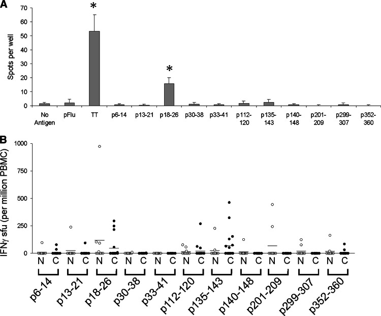
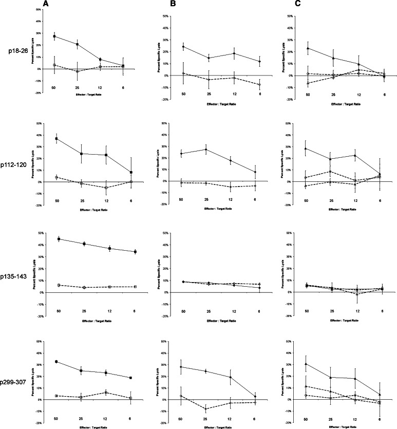
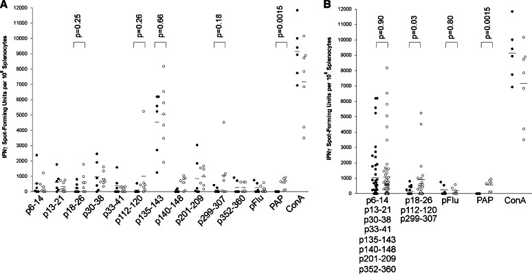
Prostatic acid phosphatase (PAP) has been investigated as the target of several antigen-specific anti-prostate tumor vaccines. The goal of antigen-specific active immunotherapies targeting PAP would ideally be to elicit PAP-specific CD8+ effector T cells. The identification of PAP-specific CD8+ T-cell epitopes should provide a means of monitoring the immunological efficacy of vaccines targeting PAP, and these epitopes might themselves be developed as vaccine antigens. In the current report, we hypothesized that PAP-specific epitopes might be identified by direct identification of pre-existing CD8+ T cells specific for HLA-A2-restricted peptides derived from PAP in the blood of HLA-A2-expressing individuals. 11 nonamer peptides derived from the amino acid sequence of PAP were used as stimulator antigens in functional ELISPOT assays with peripheral blood mononuclear cells from 20 HLA-A2+ patients with prostate cancer or ten healthy blood donors. Peptide-specific T cells were frequently identified in both groups for three of the peptides, p18-26, p112-120, and p135-143. CD8+ T-cell clones specific for three peptides, p18-26, p112-120, and p299-307, confirmed that these are HLA-A2-restricted T-cell epitopes. Moreover, HLA-A2 transgenic mice immunized with a DNA vaccine encoding PAP developed epitope-specific responses for one or more of these three peptide epitopes. We propose that this method to first identify epitopes for which there are pre-existing epitope-specific T cells could be used to prioritize MHC class I-specific epitopes for other antigens. In addition, we propose that the epitopes identified here could be used to monitor immune responses in HLA-A2+ patients receiving vaccines targeting PAP to identify potentially therapeutic immune responses.


Figures

Fig. 1
Fig. 1
PAP-derived peptide-specific T cells identified in HLA-A2-expressing individuals by ELISPOT. a Representative ELISPOT assay using peripheral blood mononuclear cells (PBMC) from a 49-year-old patient with stage D2 prostate cancer. PBMC were stimulated once in vitro with 10 μg/ml of the indicated peptides or tetanus toxoid protein (1 LFU/ml, as a positive control), and assayed for peptide-specific IFNγ release upon restimulation with peptide, as described. In this assay, IFNγ spots in response to tetanus toxoid stimulation and PAP peptide p18–26 were highly significant (P < 0.001) compared with media only (no antigen). b ELISPOT assays were performed in identical fashion using PBMC from multiple individuals. T-cell frequencies were calculated as spot-forming units (sfu) per 106 PBMC. Open circles (N) show data for the ten volunteer blood donors (n = 10), and closed circles (C) show data for the patients with prostate cancer (n = 20)
Fig. 2
Fig. 2
Peptide-specific T-cell clones specific for p18–26, p112–120, and p299–307, but not p135–143, can lyse PAP-expressing cells in an HLA-A2-restricted fashion. Clonal T-cell lines specific for p18–26, p112–120, p135–143, or p299–307 were assessed for their ability to lyse peptide-pulsed T2 target cell lines (column a), HLA-A2-expressing TK6 cell lines expressing PAP (TK6-PAP) versus control TK6 cells expressing GFP (TK6-GFP, column b), or prostate cancer cell lines (column c). Cytotoxicity was measured as LDH release after 4-h co-culture of effector cells with target cells. Assays were performed in triplicate at multiple effector-to-target ratios as indicated. Shown is the mean and standard deviation of percent specific lysis at each effector-to-target ratio, determined from triplicate assessments, of each clonal line and target cell line. column a: solid squares represent T2 cells pulsed with the relevant peptide, and open squares represent T2 target cells pulsed with an irrelevant HLA-A2-binding peptide. column b: solid diamonds represent TK6-PAP target cells, and open diamonds represent TK6-GFP target cells. column c: closed triangles represent HLA-A2-transduced LNCaP target cells (expressing PAP), and open triangles represent HLA-A2-transduced LNCaP target cells in the presence of an HLA-A2 blocking antibody. Open circles represent HLA-A2-transduced DU145 target cells (not expressing PAP). Results are representative of multiple independent experiments
Fig. 3
Fig. 3
PAP-specific epitopes can be used for monitoring immune responses following DNA immunization. HHD-II mice were immunized six times at 14-day intervals with plasmid DNA encoding PAP (pTVG-HP, n = 7) or vector control (pTVG4, n = 6). Splenocytes obtained 2 weeks after the final immunization were cultured with all peptides in bulk and assessed after 7 days by IFNγ ELISPOT using individual peptides, an HLA-A2-specific influenza control peptide (pFlu), PAP protein (PAP), or concanavalin A (ConA) as stimulatory antigens. The frequency of IFNγ sfu per 106 is shown for each animal. Closed circles represent data from vector control-immunized mice, and open circles represent data from PAP DNA-immunized mice. Comparisons of means (shown as lines) between groups are made with an unpaired t test, and the P values are shown for the relevant comparisons. a Analysis of IFNγ ELISPOT responses by individual stimulatory peptide antigens. b The same data but pooled with respect to the PAP-derived non-epitope peptides or PAP-specific HLA-A2 epitopes

Similar articles

Cited by

References

    1. Gutman AB, Gutman EB. An acid phosphatase in the serum of patients with metastasizing carcinoma of the prostate gland. J Clin Invest. 1938;17:473–479. doi: 10.1172/JCI100974. - DOI - PMC - PubMed
    1. Jobsis AC, De Vries GP, Anholt RR, Sanders GT. Demonstration of the prostatic origin of metastases: an immunohistochemical method for formalin-fixed embedded tissue. Cancer. 1978;41:1788–1793. doi: 10.1002/1097-0142(197805)41:5<1788::AID-CNCR2820410521>3.0.CO;2-D. - DOI - PubMed
    1. Siddall JK, Cooper EH, Newling DWW, Robinson MRG, Whelan P. An evaluation of the immunochemical measurement of prostatic acid phosphatase and prostatic specific antigen in carcinoma of the prostate. Eur Urol. 1986;12:123–130. - PubMed
    1. O’Keefe DS, Bacich DJ, Heston WD. Comparative analysis of prostate-specific membrane antigen (PSMA) versus a prostate-specific membrane antigen-like gene. Prostate. 2004;58:200–210. doi: 10.1002/pros.10319. - DOI - PubMed
    1. Laus R, Yang DM, Ruegg CL, Shapero MH, Slagle PH, Small E, Burch P, Valone FH. Dendritic cell immunotherapy of prostate cancer: preclinical models and early clinical experience. Canc Res Ther Control. 2001;11:1–10.

Publication types

MeSH terms