. 2010 Jul;30(5):743-50.
doi: 10.1007/s10571-010-9499-7. Epub 2010 Feb 6.

Dopaminergic modulation of spiny neurons in the turtle striatum

Affiliations

Dopaminergic modulation of spiny neurons in the turtle striatum

Jaime Barral et al. Cell Mol Neurobiol. 2010 Jul.

Abstract

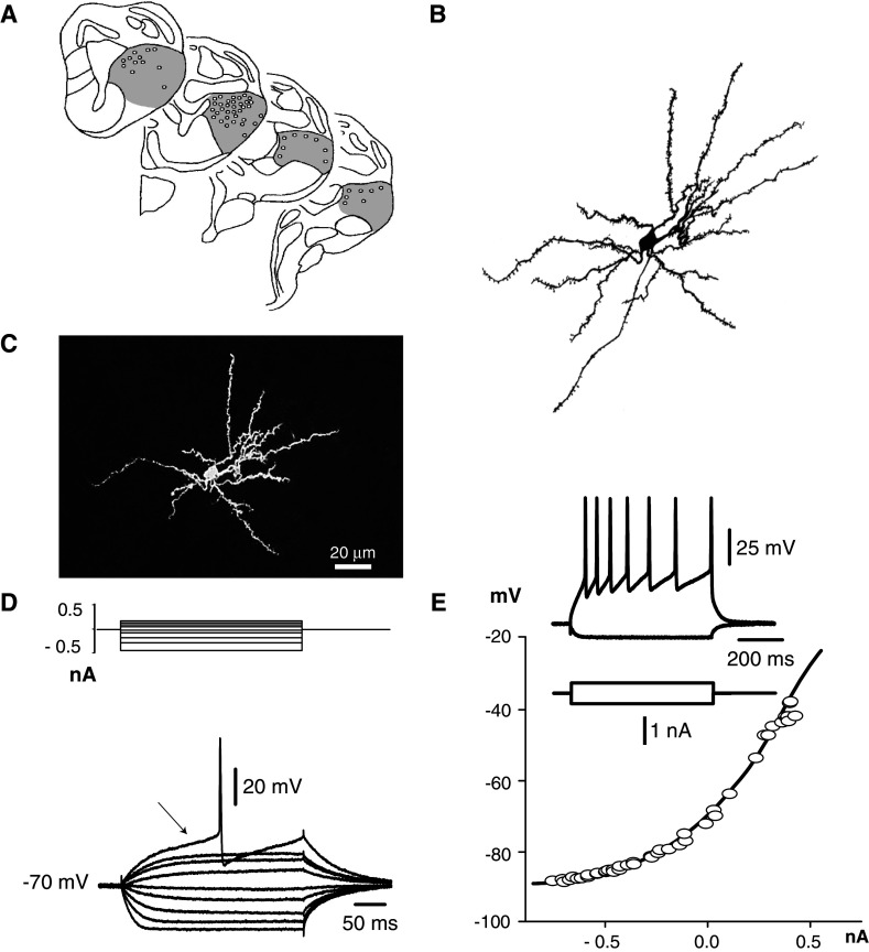
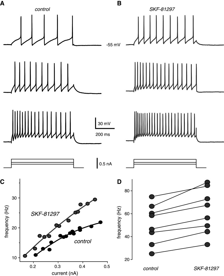
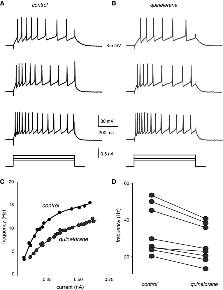
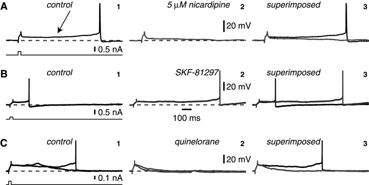
Intracellular recordings were obtained from brain slice preparation in neurons of the striatum of the turtle Trachemys scripta elegans, analogous to the mammalian striatum in its topographic organization, synaptic connectivity, cytoarchitecture, and neurochemistry. Here we show that these similarities extend to the electrophysiological properties of its neurons. Biocytin staining revealed that 85% of the recorded neurons were medium spiny neurons while 15% were aspiny neurons. Spiny neurons of the turtle resembled those found in the mammalian and avian striatum and express dopaminergic D(1) and D(2) class receptors. Because the striatum of the turtle receives a dense dopaminergic innervation from tegmental dopaminergic neurons we investigated the postsynaptic actions of selective dopamine receptor agonists in the excitability of spiny neurons. As in mammals and birds, activation of D(1)-receptors enhances, whereas activation of D(2)-receptors decreases the evoked discharge. Apparently, actions of dopamine agonists occur via the modulation of L-type (Ca(V)1) Ca2+-conductances. Strong cellular evidence suggests that the role of dopamine in the modulation of motor networks is preserved along vertebrate evolution.


Figures

Fig. 1
Fig. 1
Medium spiny neurons of the striatum of the turtle. a Coronal sections through the turtle’s brain are shown schematically. Grey regions indicate the striatum (modified from Powers and Reiner, 1980) and dots show location and distribution of recorded neurons. b Camera lucida serial reconstruction of a recorded and biocytin-filled medium spiny neuron and c photomicrograph of the same neuron. d Top traces show injected depolarizing and hyperpolarizing current steps and bottom shows voltage responses to current pulse injections to the medium spiny neuron shown in b, c. Ramp-like increase in membrane potential and latency for first action potential firing (arrow) characterize these neurons. e Current–voltage relationship (IV plot) measured from traces similar to those in d (empty circles) exhibits inward rectification. Line is a third-order polynomial fit to the data. Inset shows tonic-evoked discharge with frequency adaptation
Fig. 2
Fig. 2
Activation of dopamine D1-class receptors enhances the excitability of spiny neurons in the striatum of the turtle. a Control: repetitive discharge evoked by depolarizing rectangular current steps of increasing strength (at the bottom). b Evoked discharge after bath application of the selective dopamine D1-receptor agonist SKF-81297 (1 μM). Firing frequency to the same current steps was increased. c Intensity–frequency relationships (IF plots) built from traces as those in ab. Note increased gain for similar stimuli after the D1-agonist. d Paired line plot showing variation in mean frequency (measured at half maximum frequency in IF plots) after addition of the D1-agonist
Fig. 3
Fig. 3
Activation of dopamine D2-class receptors depresses the excitability of spiny neurons in the striatum of the turtle. a Control: repetitive discharge evoked by depolarizing rectangular current steps of increasing strength (at the bottom). b Evoked discharge after bath application of the selective dopamine D2-receptor agonist quinelorane (1 μM). Firing frequency to the same current steps was decreased. c Intensity–frequency relationships (IF plots) built from traces as those in ab. Note decreased gain for similar stimuli after the D2-agonist. d Paired line plot showing variation in mean frequency (measured at half maximum frequency in IF plots) after addition of the D2-agonist
Fig. 4
Fig. 4
Subthreshold action of dopamine agonists in the striatum of the turtle. a As it is the case of mammalian striatal neurons (Hernández-López et al. 1997), spiny neurons in the striatum of the turtle may respond with prolonged subthreshold depolarizations (arrow at the control) to brief current stimulus (at the bottom) if given at a depolarized membrane potential (≈ −50 mV) (1). These depolarizations may last hundreds of milliseconds and may produce the firing of action potentials much time after the stimulus is over. In the turtle as in the rodent, dihydropyridines (5 μM nicardipine in this case) reduce the subthreshold response (2). Superimposed traces are shown in 3. b In neurons in which the subthreshold depolarization is not very prolonged (control in 1), bath applications of the dopaminergic D1-class agonist SKF81297 (1 μM) (2) prolong the slow subthreshold response. 1 and 2 are superimposed in 3. c On the contrary, the dopaminergic D2-class receptor agonist quinelorane (5 μM) reduces the duration of the subthreshold response and may abolish neuronal firing altogether (cf., 1 and 2)

Similar articles

Cited by

References

    1. Andersen A, Baestrup C, Randrup A (1975) Apomorphine-induced stereotyped biting in the tortoise in relation to dopaminergic mechanisms. Brain Behav Evol 11:365–373 - PubMed
    1. Bargas J, Galarraga E, Aceves J (1989) An early outward conductance modulates the firing latency and frequency of neostriatal neurons of the rat brain. Exp Brain Res 75:146–156 - PubMed
    1. Brauth SE, Reiner A, Kitt CA, Karten HJ (1983) The substance P-containing striato-tegmental path in reptiles: an immunohistochemical study. J Comp Neurol 219:305–327 - PubMed
    1. Butler A, Hodos W (2005). Comparative vertebrate neuroanatomy, evolution and adaptation, 2nd edn. Wiley Interscience, p 484
    1. Carrillo-Reid L, Tecuapetla F, Tapia D, Hernandez-Cruz A, Galarraga E, Drucker-Colin R, Bargas J (2008) Encoding network states by striatal cell assemblies. J Neurophysiol 99:1435–1450 - PubMed

Publication types

MeSH terms

LinkOut - more resources