

Enhanced bone formation in large segmental radial defects by combining adipose-derived stem cells expressing bone morphogenetic protein 2 with nHA/RHLC/PLA scaffold
- PMID: 20140671
- PMCID: PMC2989079
- DOI: 10.1007/s00264-009-0946-3
Enhanced bone formation in large segmental radial defects by combining adipose-derived stem cells expressing bone morphogenetic protein 2 with nHA/RHLC/PLA scaffold
Abstract
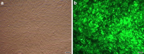
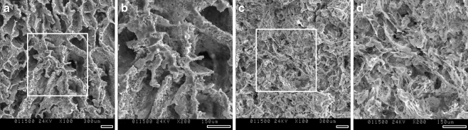
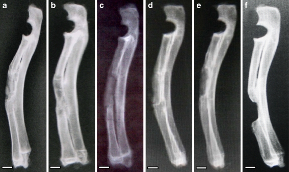
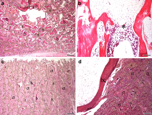
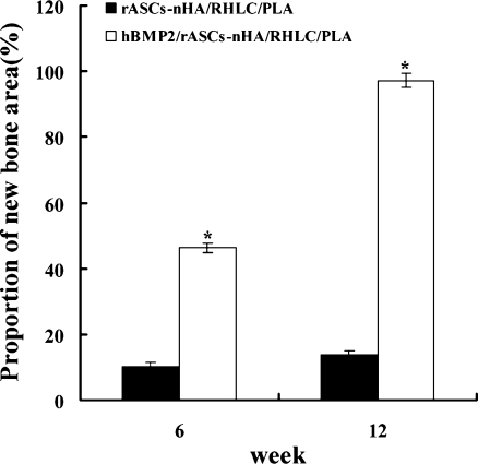
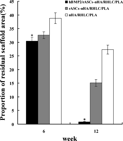
In this study, rabbit adipose-derived stem cells (rASCs) were isolated, cultured in vitro, and transfected with recombinant adenovirus vector containing human bone morphogenetic protein 2 (Ad-hBMP2). These cells were combined with a nano-hydroxyapatite/recombinant human-like collagen/poly(lactic acid) scaffold (nHA/RHLC/PLA) to fabricate a new biocomposite (hBMP2/rASCs-nHA/RHLC/PLA, group 1) and cultured in osteogenic medium. Non-transfected rASCs mixed with nHA/RHLC/PLA (rASCs-nHA/RHLC/PLA, group 2) and nHA/RHLC/PLA scaffold alone (group 3) served as controls. Scanning electron microscope (SEM) demonstrated integration of rASCs with the nHA/RHLC/PLA scaffold. Quantitative real-time RT-PCR analyses of collagen I, osteonectin, and osteopontin cDNA expression indicated that the osteogenic potency of rASCs was enhanced by transfection with Ad-hBMP2. After in vitro culture for seven days, three groups were implanted into 15-mm length critical-sized segmental radial defects in rabbits. After 12 weeks, radiographic and histological analyses were performed. In group 1, the medullary cavity was recanalised, bone was rebuilt and moulding was finished, the bone contour had begun to remodel and scaffold was degraded completely. In contrast, bone defects were not repaired in groups 2 or 3. Furthermore, the scaffold degradation rate in group 1 was significantly higher than in groups 2 or 3. In summary, after transduction with Ad-hBMP2, the osteogenesis of rASCs was enhanced; a new biocomposite created with these cells induced repair of a critical bone defect in vivo in a relatively short time.
Figures

Similar articles
-
[Experimental study of repairing femoral bone defects with nHA/RHLC/PLA scaffold composite with endothelial cells and osteoblasts in canines].Zhonghua Yi Xue Za Zhi. 2013 May 7;93(17):1335-40. Zhonghua Yi Xue Za Zhi. 2013. PMID: 24029485 Chinese.
-
Improving osteogenesis of three-dimensional porous scaffold based on mineralized recombinant human-like collagen via mussel-inspired polydopamine and effective immobilization of BMP-2-derived peptide.Colloids Surf B Biointerfaces. 2017 Apr 1;152:124-132. doi: 10.1016/j.colsurfb.2016.12.041. Epub 2017 Jan 4. Colloids Surf B Biointerfaces. 2017. PMID: 28103529
-
Skeletal repair in rabbits using a novel biomimetic composite based on adipose-derived stem cells encapsulated in collagen I gel with PLGA-beta-TCP scaffold.J Orthop Res. 2010 Feb;28(2):252-7. doi: 10.1002/jor.20969. J Orthop Res. 2010. PMID: 19688871
-
Nano-Hydroxyapatite as a Delivery System for Promoting Bone Regeneration In Vivo: A Systematic Review.Nanomaterials (Basel). 2021 Sep 29;11(10):2569. doi: 10.3390/nano11102569. Nanomaterials (Basel). 2021. PMID: 34685010 Free PMC article. Review.
-
Recent developments in biomaterials for long-bone segmental defect reconstruction: A narrative overview.J Orthop Translat. 2019 Oct 8;22:26-33. doi: 10.1016/j.jot.2019.09.005. eCollection 2020 May. J Orthop Translat. 2019. PMID: 32440496 Free PMC article. Review.
Cited by
-
MicroRNA-26a-modified adipose-derived stem cells incorporated with a porous hydroxyapatite scaffold improve the repair of bone defects.Mol Med Rep. 2015 Sep;12(3):3345-3350. doi: 10.3892/mmr.2015.3795. Epub 2015 May 18. Mol Med Rep. 2015. PMID: 25997460 Free PMC article.
-
Osteogenic differentiation is synergistically influenced by osteoinductive treatment and direct cell-cell contact between murine osteoblasts and mesenchymal stem cells.Int Orthop. 2012 Jan;36(1):199-205. doi: 10.1007/s00264-011-1259-x. Epub 2011 May 13. Int Orthop. 2012. PMID: 21567150 Free PMC article.
-
Smart scaffolds in bone tissue engineering: A systematic review of literature.World J Stem Cells. 2015 Apr 26;7(3):657-68. doi: 10.4252/wjsc.v7.i3.657. World J Stem Cells. 2015. PMID: 25914772 Free PMC article.
-
Partial load-bearing rabbit ulnar segmental defects are regenerated with biocompatible grafts with or without bone marrow-derived mesenchymal stem cells.Ulus Travma Acil Cerrahi Derg. 2022 Aug;28(8):1066-1072. doi: 10.14744/tjtes.2021.64569. Ulus Travma Acil Cerrahi Derg. 2022. PMID: 35920424 Free PMC article.
-
Bone Regeneration Using Bone Morphogenetic Proteins and Various Biomaterial Carriers.Materials (Basel). 2015 Apr 15;8(4):1778-1816. doi: 10.3390/ma8041778. Materials (Basel). 2015. PMID: 28788032 Free PMC article. Review.
References
MeSH terms
Substances
LinkOut - more resources
Full Text Sources
Other Literature Sources
Research Materials
