. 2010 May;67(10):1661-74.
doi: 10.1007/s00018-010-0270-5. Epub 2010 Feb 7.

Estrogen augments the T cell-dependent but not the T-independent immune response

Affiliations

Estrogen augments the T cell-dependent but not the T-independent immune response

Mónika Adori et al. Cell Mol Life Sci. 2010 May.

Erratum in

  • Cell Mol Life Sci. 2010 Jul;67(14):2509. Kövesdi, Dorottya [added]

Abstract

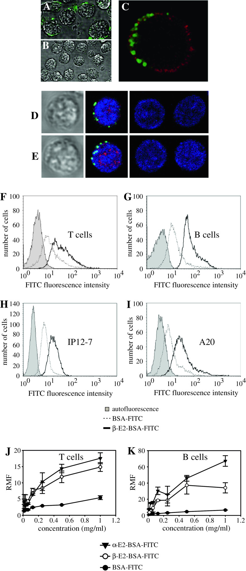
Estrogen plays a critical regulatory role in the development and maintenance of immunity. Its role in the regulation of antibody synthesis in vivo is still not completely clear. Here, we have compared the effect of estrogen on T cell-dependent (TD) and T cell-independent type 2 (TI-2) antibody responses. The results provide the first evidence that estrogen enhances the TD but not the TI-2 response. Ovariectomy significantly decreased, while estrogen re-administration increased the number of hapten-specific IgM- and IgG-producing cells in response to TD antigen. In vitro experiments also show that estrogen may have a direct impact on B and T cells by inducing rapid signaling events, such as Erk and AKT phosphorylation, cell-specific Ca(2+) signal, and NFkappaB activation. These non-transcriptional effects are mediated by classical estrogen receptors and partly by an as yet unidentified plasma membrane estrogen receptor. Such receptor- mediated rapid signals may modulate the in vivo T cell-dependent immune response.


Figures

Fig. 1
Fig. 1
Effect of ovariectomy on IgG and IgM production by mouse splenocytes following T cell-dependent or -independent immune responses showing the IgM (a) and IgG (b) contents of the sera following immunization with TD antigen (a,b, respectively) or TI-2 antigen (c,d, respectively). The results are means ± SD. Values from triplicate samples
Fig. 2
Fig. 2
Analysis of hapten specific IgG- and IgM-producing cells as a response to TD and TI-2 antigens in mice: effect of ovariectomy and estrogen replacement. The number of IgM- (a) and IgG- (b) producing cells are shown as a response to immunization with TD antigen in OVX- and SHAM-operated control mice. The same cell numbers in response to TI-2 antigen are shown in c,d, respectively. The numbers of IgM- and IgG-producing cells (e,f, respectively) are displayed in OVX mice immunized with TD antigen under estrogen replacement conditions. The results are means ± SD of triplicate samples from multiple groups of mice. Significance analysis was performed as described in “Materials and methods”
Fig. 3
Fig. 3
Classical estrogen receptors, ERα and ERβ, are expressed in murine B and T lymphocytes. ERα and ERβ receptors are expressed in both primary murine lymphocytes (a right panels) and in T and B cell lines (a left panels) as assessed by flow cytometry (a) microscopy (b upper panel). Representative confocal microscopic images show expression and mainly cytoplasmic localization of both ERα and ERβ in both B and T splenic lymphocytes (b upper panels). Upon βE2 treatment, significant nuclear translocation of ERα is observed (b lower right) (red cholera toxin B membrane staining; green intracellular staining with anti-ERα; blue nuclear counterstaining). The presence of ERα and ERβ proteins in T and B cell lysates was also analyzed by western blots (b lower left). Anti-SHP-2 was used as loading control, and, to see the specificity of the signal, only the secondary antibody (goat anti-rabbit IgG-HRPO) was added (b bottom left)
Fig. 4
Fig. 4
Binding of cell-impermeant βE2-BSA conjugates to B and T cells. Representative confocal microscopic image (DIC + fluorescence overlay) shows cell surface binding of 17-β-E2-BSA-FITC to splenic T lymphocytes (a), while BSA-FITC showed no significant binding (b). Similar patchy membrane staining was also observed in B lymphocytes (c) (green 17-β-E2-BSA-FITC; red anti-B220 antibody). This was quantitatively confirmed by flow cytometry in both primary lymphocytes and the T and B cell lines (fi). Representative images of T cells (d) and B cells (e) double stained with 17-β-E2-BSA-FITC (green) and anti-ERα (red) with nuclear counterstaining (blue) show disparate staining for ERα and the membrane βE2 binding site. j,k Dose-dependence of 17-β- and 17-α-E2-BSA-FITC binding (measured 15 min after addition) to T cells and B cells, respectively. As a control, BSA-FITC binding is also shown. Error bars SD calculated from three independent experiments. RMF Relative mean fluorescence intensity, normalized to autofluorescence
Fig. 5
Fig. 5
Calcium signals induced by estrogen in splenic B and T lymphocytes. Fluo-4 loaded murine splenocytes (a,b), purified B splenocytes (c,e) and T splenocytes (d,f) were investigated by microscopy for their single cell Ca2+ response to 100 nM membrane permeable or impermeable 17β-estradiol (βE2 and βE2-BSA, respectively). Representative single cell recordings of three individual cells demonstrate the substantial heterogeneity among splenocytes in their estradiol-induced Ca2+ response (a). b The averaged response of splenocytes (n = 58) to βE2. In separated murine B splenocytes, neither βE2 nor βE2-BSA cause significant changes in the intracellular Ca2+ level (c,e) (n = 27 and n = 26, respectively), while purified T cells responded to both forms of estradiol (d,f) (n = 54 and n = 55, respectively). The “maximal response” of the cells to ionomycin Ca2+ ionophore is also shown in cf (right side)
Fig. 6
Fig. 6
Estrogen signals modulate ERK and Akt phosphorylation of B and T lymphocytes. Separated murine B and T splenocytes were treated with βE2 (a) or with 17-β-E2-BSA (b). Cell lysates were examined for ERK and Akt phosphorylation by western blot. a The B and T cells were stimulated, as a positive control, with anti-IgM and anti-CD3, respectively, and with 17-β-estradiol for different times as indicated under the lanes. The lower panels demonstrate densitometric and the statistical evaluation of the WBs. This evaluation based on relative density values of the bands normalized to total SHP-2 level, as described in “Materials and methods”. b The same western blot analysis is shown for 17-β-estradiol-BSA conjugate. c Analysis of the effects by 17-α- estradiol-BSA conjugate on T and B cells is shown under the same conditions as in (a) and (b)
Fig. 7
Fig. 7
Estrogen induces NFκB activation/nuclear translocation and IFNγ gene activation in splenic B and T cells. a Representative (from 150 cells) DIC and fluorescence images of control and βE2-treated murine splenic T and B cells stained for p65 NF-κB (green) and DNA/nucleus (red). b Nuclear translocation of cRel, RelA (p65), and p100/p52 NF-κB proteins in purified murine splenic B cells as assessed by immunoblotting. Cells were left untreated (control) or treated with anti-IgM, 10 nM 17-β-E2, and vehicle (ethanol) for 60 min. c IFNγ levels were determined from supernatants of 72-h cell cultures of unseparated splenocytes stimulated with concanavalin A (ConA) alone, or with 1 and 10 nM 17-β-E2 by ELISA. The results are means ± SD of three independent cultures

References

    1. Tanriverdi F, Silveira LF, MacColl GS, Bouloux PM. The hypothalamic-pituitary-gonadal axis: immune function and autoimmunity. J Endocrinol. 2003;176:293–304. doi: 10.1677/joe.0.1760293. - DOI - PubMed
    1. Ansar AS, Penhale WJ, Talal N. Sex hormones, immune responses, and autoimmune diseases. Mechanisms of sex hormone action. Am J Pathol. 1985;121:531–551. - PMC - PubMed
    1. Lang TJ. Estrogen as an immunomodulator. Clin Immunol. 2004;113:224–230. doi: 10.1016/j.clim.2004.05.011. - DOI - PubMed
    1. Grimaldi CM, Hicks R, Diamond B. B cell selection and susceptibility to autoimmunity. J Immunol. 2005;174:1775–1781. - PubMed
    1. Grimaldi CM, Cleary J, Dagtas AS, Moussai D, Diamond B. Estrogen alters thresholds for B cell apoptosis and activation. J Clin Invest. 2002;109:1625–1633. - PMC - PubMed

Publication types

MeSH terms

LinkOut - more resources