Review
. 2010 May;67(9):1423-45.
doi: 10.1007/s00018-010-0268-z. Epub 2010 Feb 7.

From carrot to clinic: an overview of the retinoic acid signaling pathway

Affiliations
Review

From carrot to clinic: an overview of the retinoic acid signaling pathway

Maria Theodosiou et al. Cell Mol Life Sci. 2010 May.

Erratum in

  • Cell Mol Life Sci. 2010 May;67(9):1563-4

Abstract

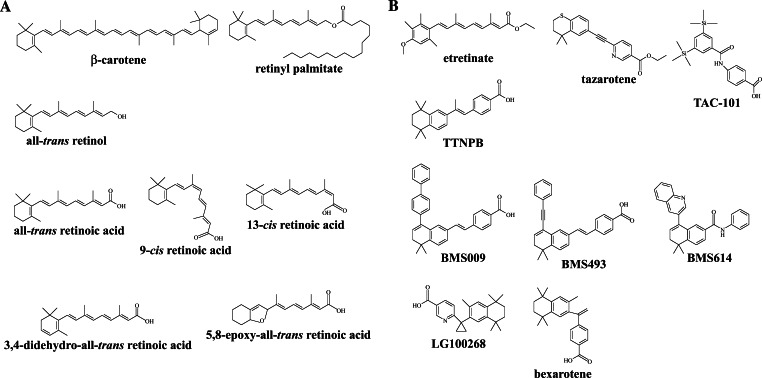
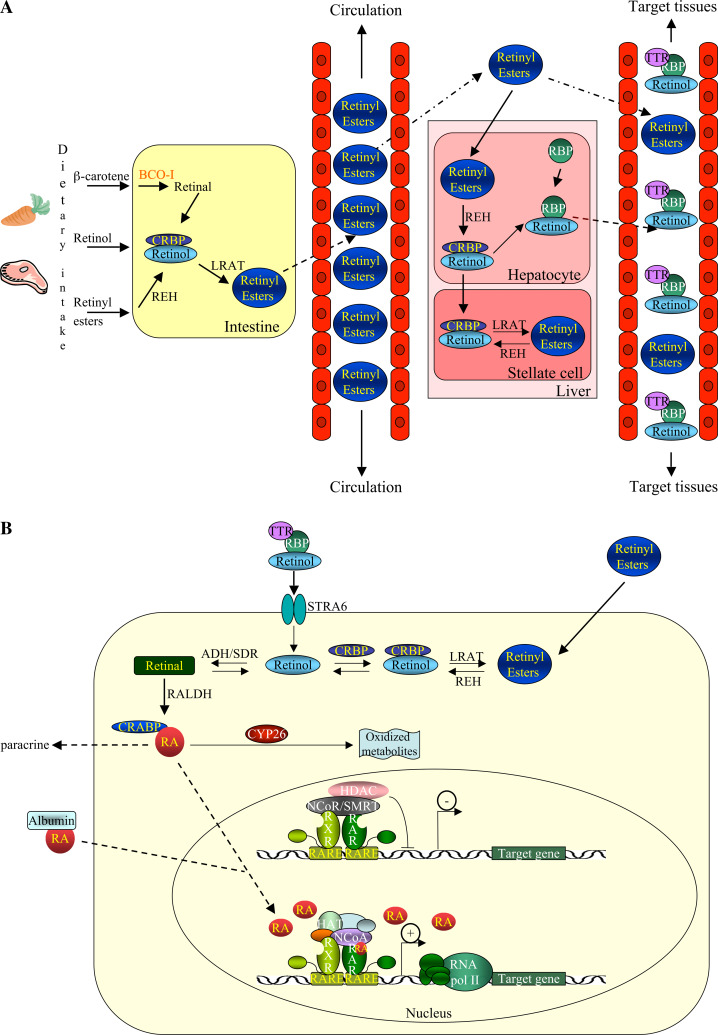
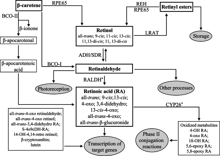
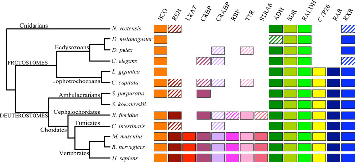
Vitamin A is essential for the formation and maintenance of many body tissues. It is also important for embryonic growth and development and can act as a teratogen at critical periods of development. Retinoic acid (RA) is the biologically active form of vitamin A and its signaling is mediated by the RA and retinoid X receptors. In addition to its role as an important molecule during development, RA has also been implicated in clinical applications, both as a potential anti-tumor agent as well as for the treatment of skin diseases. This review presents an overview of how dietary retinoids are converted to RA, hence presenting the major players in RA metabolism and signaling, and highlights examples of treatment applications of retinoids. Moreover, we discuss the origin and diversification of the retinoid pathway, which are important factors for understanding the evolution of ligand-specificity among retinoid receptors.


Figures

Fig. 1
Fig. 1
Natural and synthetic retinoids. a Structures of natural retinoids and of their catabolism products. b Synthetic agonists and antagonists of RAR and RXR
Fig. 2
Fig. 2
Metabolism of vitamin A and transcriptional activation. a Dietary intake of retinoids and absorption in the intestine. In the intestine, retinoids are converted to retinol, which is bound by CRBP. This retinol is further processed to retinyl esters and exported into the circulation, where the retinyl esters are taken to the liver. In hepatocytes of the liver, retinyl esters are converted to retinol and bound to RBP for transport to target cells. In stellate cells of the liver, retinol is converted to retinyl esters for storage. In the bloodstream, the retinol:RBP complex is bound to TTR to avoid elimination by the kidney and to ensure delivery to target tissues. b Retinol is delivered to target cells in a complex with RBP and TTR and uptake occurs via the STRA6 receptor. In the target cell, free retinol is oxidized to retinaldehyde by ADH/SDR enzymes in a reversible reaction. A second, irreversible oxidation is catalyzed by RALDH and converts retinaldehyde to RA, which is bound by CRABP. RA enters the nucleus, where RAR/RXR heterodimers are bound to RAREs and associated with a co-repressor complex. Binding of RA induces a conformational change of the RAR/RXR heterodimer that results in release of co-repressors and recruitment of co-activators and initiation of transcription. RA is degraded and eliminated by CYP26 enzymes. ADH Alcohol dehydrogenase, BCO-I β,β-carotene-15,15′-monooxygenase, CRABP cellular retinoic acid binding protein, CRBP cellular retinol binding protein, CYP26 cytochrome P450 family 26, HAT histone acetyltransferase, HDAC histone deacetylase, LRAT lecithin:retinol acetyltransferase, NCoA co-activator complex, NCoR co-repressor complex, RA retinoic acid, RALDH retinaldehyde dehydrogenase, RAR retinoic acid receptor, RARE retinoic acid response element, RBP retinol binding protein, REH retinyl ester hydrolase, RNA pol II ribonucleic acid polymerase II, RXR retinoid X receptor, SDR short-chain dehydrogenase/reductase, SMRT silencing mediator of retinoic acid and thyroid hormone receptor, STRA6 stimulated by retinoic acid gene 6, TTR transthyretin
Fig. 3
Fig. 3
Biochemical pathway of retinoids. Endogenous retinoids are boxed and the major physiological endpoints of the metabolic pathway are indicated in gray circles. The enzymes responsible for conversion of the retinoids are indicated. The asterisks indicate that certain enzymes of the CYP family can catalyze the synthesis or degradation of RA in vitro. ADH Alcohol dehydrogenase (ADH1, 3, 4), BCO-I β,β-carotene-15,15′-monooxygenase, BCO-II β,β-carotene-9′,10′-dioxygenase, CYP26 cytochrome P450 family 26, LRAT lecithin:retinol acetyltransferase, RA retinoic acid, RALDH retinaldehyde dehydrogenases (RALDH1, 2, 3, 4), REH retinol ester hydrolase, RPE65 retinal pigment epithelium-specific protein 65 kDa, SDR short-chain dehydrogenase/reductase
Fig. 4
Fig. 4
Molecular components of RA metabolism and signaling in different metazoans. Using vertebrate sequences for each component, the genomes of the respective species were searched using BLAST. The best hits were then used for reverse BLAST searches of the respective vertebrate genomes to verify the association of a given sequence with the protein family of the RA signaling component in question. Hatched boxes indicate that representatives of a given protein family might exist in a given animal group, but that our BLAST analyses were not conclusive. For example, for CRBP and CRABP, the hatched boxes indicate that members of the fatty acid binding protein family have been identified, and, for RXR, the hatched boxes highlight that this protein is absent from the assayed species, but present in other members of this phylum. The species are: Branchiostoma floridae (amphioxus), Caenorhabditis elegans (nematode worm), Capitella capitata (annelid worm), Ciona intestinalis (sea squirt), Daphnia pulex (water flea), Drosophila melanogaster (fruit fly), Homo sapiens (human), Lottia gigantea (gastropod snail), Mus musculus (mouse), Nematostella vectensis (sea anemone), Rattus norvegicus (rat), Saccoglossus kowalevskii (acorn worm), Strongylocentrotus purpuratus (sea urchin). ADH Alcohol dehydrogenase (ADH1, 3, 4), BCO BCO-I (β,β-carotene-15,15′-monooxygenase) or BCO-II (β,β-carotene-9′,10′-dioxygenase) or RPE65 (retinal pigment epithelium-specific protein 65 kDa), CRABP cellular retinoic acid binding protein, CRBP cellular retinol binding protein, CYP26 cytochrome P450 family 26, LRAT lecithin:retinol acetyltransferase, RALDH retinaldehyde dehydrogenases (RALDH1, 2, 3, 4), RAR retinoic acid receptor, RBP retinol binding protein, REH retinol ester hydrolase, RXR retinoid X receptor, SDR short-chain dehydrogenase/reductase, STRA6 stimulated by retinoic acid gene 6, TTR transthyretin

Similar articles

Cited by

References

    1. Collins MD, Mao GE. Teratology of retinoids. Annu Rev Pharmacol Toxicol. 1999;39:399–430. doi: 10.1146/annurev.pharmtox.39.1.399. - DOI - PubMed
    1. Morriss-Kay GM, Ward SJ. Retinoids and mammalian development. Int Rev Cytol. 1999;188:73–131. doi: 10.1016/S0074-7696(08)61566-1. - DOI - PubMed
    1. Kastner P, Mark M, Chambon P. Nonsteroid nuclear receptors: what are genetic studies telling us about their role in real life? Cell. 1995;83:859–869. doi: 10.1016/0092-8674(95)90202-3. - DOI - PubMed
    1. Blomhoff R, Blomhoff HK. Overview of retinoid metabolism and function. J Neurobiol. 2006;66:606–630. doi: 10.1002/neu.20242. - DOI - PubMed
    1. Sommer A. Xerophthalmia and vitamin A status. Prog Retin Eye Res. 1998;17:9–31. doi: 10.1016/S1350-9462(97)00001-3. - DOI - PubMed

Publication types

MeSH terms

LinkOut - more resources