

Divergent regulation of angiopoietin-1 and -2, Tie-2, and thrombospondin-1 expression by estrogen in the baboon endometrium
- PMID: 20140967
- PMCID: PMC2852108
- DOI: 10.1002/mrd.21163
Divergent regulation of angiopoietin-1 and -2, Tie-2, and thrombospondin-1 expression by estrogen in the baboon endometrium
Abstract
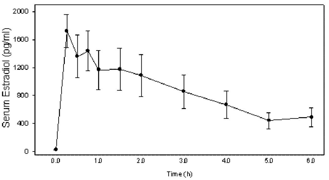
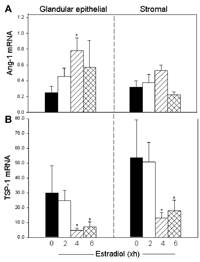
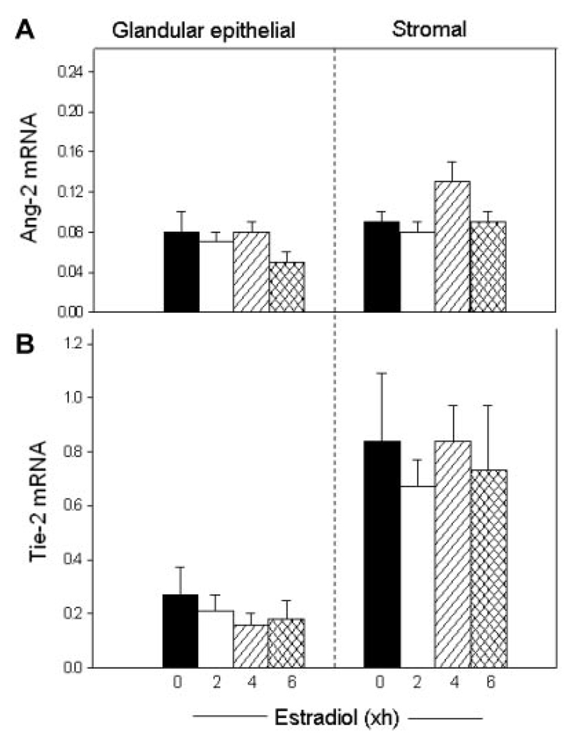
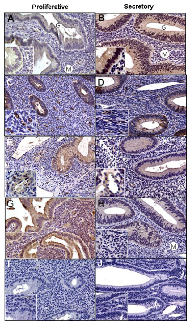
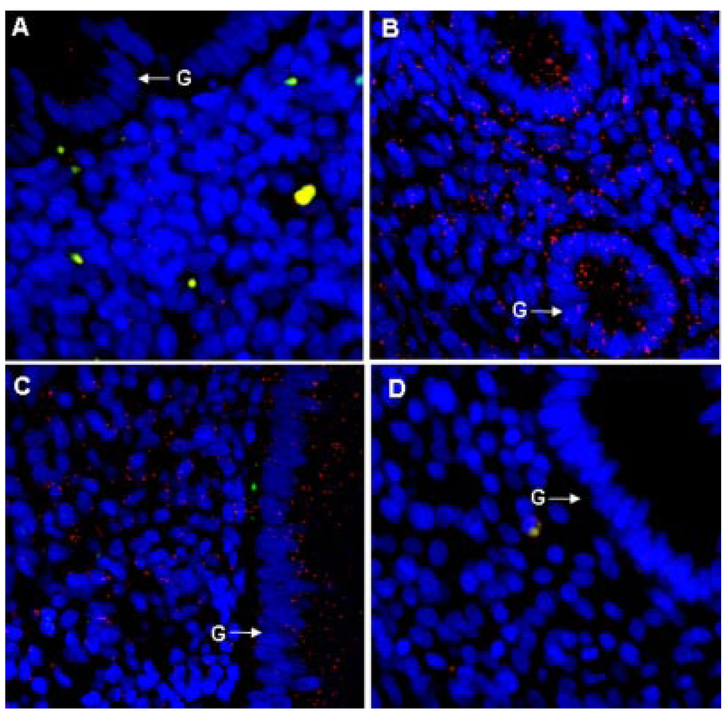
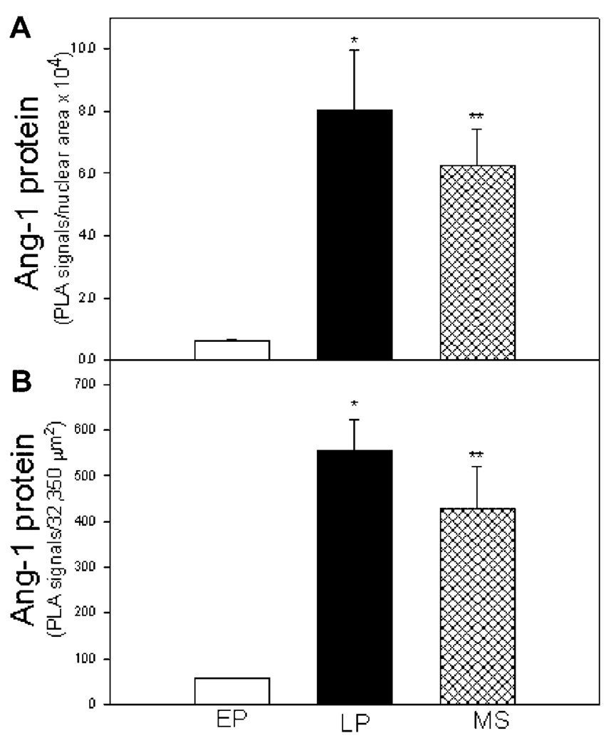
Estrogen has an important role in the reconstruction of a new vascular network in the endometrium during each menstrual cycle; however, the underlying mechanisms are incompletely understood. Angiopoietin-1 (Ang-1) promotes vessel assembly, whereas Ang-2 and thrombospondin-1 (TSP-1) cause vessel breakdown. To determine the potential effect of estrogen on the expression of these angioregulatory factors in the endometrium, Ang-1, Ang-2, TSP-1, and Tie-2 receptor mRNA levels were assessed by real-time reverse transcriptase polymerase chain reaction in glandular epithelial and stromal cells isolated from the endometrium of ovariectomized baboons treated acutely with estradiol. Corresponding protein expression was assessed by immunocytochemistry and the proximity ligation assay (PLA) during advancing stages of the baboon menstrual cycle. Serum estradiol levels in ovariectomized baboons were 400 pg/ml within 4-6 hr of estradiol treatment. Ang-1 mRNA levels in glandular epithelial cells increased threefold (P < 0.01) within 4 hr of estradiol administration. In contrast, TSP-1 mRNA levels decreased four- to fivefold (P < 0.01) in endometrial glandular epithelial and stromal cells 4-6 hr after estradiol, whereas Ang-2 and Tie-2 expression was unaltered. Immunostaining for Ang-1 increased, TSP-1 decreased, and Ang-2 and Tie-2 were unaltered in the endometrium during the secretory compared with the proliferative phase of the cycle. Endometrial Ang-1 protein expression, quantified by PLA, increased 10-fold (P < 0.05) between the early proliferative and late proliferative/mid-secretory phases of the menstrual cycle in association with the rise in estrogen. In summary, estrogen induced a rapid, divergent, and cell-specific change in expression of angiostimulatory and angioinhibitory growth factors in the endometrium of the nonhuman primate.
Figures
Similar articles
-
Effect of estrogen on vascular endothelial growth/permeability factor expression by glandular epithelial and stromal cells in the baboon endometrium.Biol Reprod. 2003 Jun;68(6):1997-2004. doi: 10.1095/biolreprod.102.011288. Epub 2003 Jan 8. Biol Reprod. 2003. PMID: 12606344
-
Expression of vascular endothelial growth/permeability factor by endometrial glandular epithelial and stromal cells in baboons during the menstrual cycle and after ovariectomy.Endocrinology. 2002 Oct;143(10):4007-17. doi: 10.1210/en.2002-220385. Endocrinology. 2002. PMID: 12239112
-
Differential expression of angiopoietins 1 and 2 and their receptor Tie-2 in human endometrium.Mol Hum Reprod. 2003 Nov;9(11):663-9. doi: 10.1093/molehr/gag083. Mol Hum Reprod. 2003. PMID: 14561809
-
Hormonal regulation and localization of estrogen, progestin and androgen receptors in the endometrium of nonhuman primates: effects of progesterone receptor antagonists.Arch Histol Cytol. 2004 Dec;67(5):393-409. doi: 10.1679/aohc.67.393. Arch Histol Cytol. 2004. PMID: 15781981 Review.
-
Secretory proteins of the baboon (Papio anubis) endometrium: regulation during the menstrual cycle and early pregnancy.Hum Reprod Update. 1997 Nov-Dec;3(6):553-9. doi: 10.1093/humupd/3.6.553. Hum Reprod Update. 1997. PMID: 9584945 Review.
Cited by
-
Angiopoietin-2 promotes ER+ breast cancer cell survival in bone marrow niche.Endocr Relat Cancer. 2016 Aug;23(8):609-23. doi: 10.1530/ERC-16-0086. Epub 2016 Jun 27. Endocr Relat Cancer. 2016. PMID: 27353038 Free PMC article.
-
Hippo Signaling in the Endometrium.Int J Mol Sci. 2022 Mar 31;23(7):3852. doi: 10.3390/ijms23073852. Int J Mol Sci. 2022. PMID: 35409214 Free PMC article. Review.
-
Female sex protects against renal edema, but not lung edema, in mice with partial deletion of the endothelial barrier regulator Tie2 compared to male sex.PLoS One. 2023 Nov 16;18(11):e0293673. doi: 10.1371/journal.pone.0293673. eCollection 2023. PLoS One. 2023. PMID: 37972011 Free PMC article.
-
Quantification of Protein Expression by Proximity Ligation Assay in the Nonhuman Primate in Response to Estrogen.Methods Mol Biol. 2022;2418:77-93. doi: 10.1007/978-1-0716-1920-9_6. Methods Mol Biol. 2022. PMID: 35119661 Free PMC article.
-
The role of decidual cells in uterine hemostasis, menstruation, inflammation, adverse pregnancy outcomes and abnormal uterine bleeding.Hum Reprod Update. 2016 Jun;22(4):497-515. doi: 10.1093/humupd/dmw004. Epub 2016 Feb 23. Hum Reprod Update. 2016. PMID: 26912000 Free PMC article. Review.
References
-
- Aberdeen GW, Wiegand SJ, Bonagura TW, Jr, Pepe GW, Albrecht ED. Vascular endothelial growth factor mediates the estrogen-induced breakdown of tight junctions between and increase in proliferation of microvessel endothelial cells in the baboon endometrium. Endocrinology. 2008;149:6076–6083. - PMC - PubMed
-
- Albrecht ED, Aberdeen GW, Niklaus AL, Babischkin JS, Suresch DL, Pepe GJ. Acute temporal regulation of vascular endothelial growth/permeability factor expression and endothelial morphology in the baboon endometrium by ovarian steroids. J Clin Endocrinol Metab. 2003;88:2844–2852. - PubMed
-
- Albrecht ED, Aberdeen GW, Pepe GJ. The role of estrogen in the maintenance of primate pregnancy. Am J Obstet Gynecol. 2000;182:432–438. - PubMed
-
- Albrecht ED, Haskins AL, Hodgen GD, Pepe GJ. Luteal function in baboons with administration of the antiestrogen ethamoxytriphetol (MER-25) throughout the luteal phase of the menstrual cycle. Biol Reprod. 1981;25:451–457. - PubMed
-
- Albrecht ED, Pepe GJ. Steroid hormone regulation of angiogenesis in the primate endometrium. Front Biosci. 2003;8:d416–d429. - PubMed
Publication types
MeSH terms
Substances
Grants and funding
LinkOut - more resources
Full Text Sources
Miscellaneous
