Electronic signatures of all four DNA nucleosides in a tunneling gap
- PMID: 20141183
- PMCID: PMC2836180
- DOI: 10.1021/nl1001185
Electronic signatures of all four DNA nucleosides in a tunneling gap
Abstract
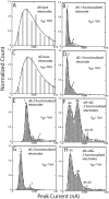
Nucleosides diffusing through a 2 nm electron-tunneling junction generate current spikes of sub-millisecond duration with a broad distribution of peak currents. This distribution narrows 10-fold when one of the electrodes is functionalized with a reagent that traps nucleosides in a specific orientation with hydrogen bonds. Functionalizing the second electrode reduces contact resistance to the nucleosides, allowing them to be identified via their peak currents according to deoxyadenosine > deoxycytidine > deoxyguanosine > thymidine, in agreement with the order predicted by a density functional calculation.
Figures


Similar articles
-
Interaction of low-energy electrons with the purine bases, nucleosides, and nucleotides of DNA.J Chem Phys. 2006 Dec 28;125(24):244302. doi: 10.1063/1.2424456. J Chem Phys. 2006. PMID: 17199346
-
Sugar pucker modulates the cross-correlated relaxation rates across the glycosidic bond in DNA.J Am Chem Soc. 2005 Oct 26;127(42):14663-7. doi: 10.1021/ja050894t. J Am Chem Soc. 2005. PMID: 16231919
-
Determination of DNA-base orientation on carbon nanotubes through directional optical absorbance.Nano Lett. 2007 Aug;7(8):2312-6. doi: 10.1021/nl070953w. Epub 2007 Jul 3. Nano Lett. 2007. PMID: 17608443
-
Recognition tunneling.Nanotechnology. 2010 Jul 2;21(26):262001. doi: 10.1088/0957-4484/21/26/262001. Epub 2010 Jun 4. Nanotechnology. 2010. PMID: 20522930 Free PMC article. Review.
-
Molecular signatures in the transport properties of molecular wire junctions: what makes a junction "molecular"?Small. 2006 Feb;2(2):172-81. doi: 10.1002/smll.200500201. Small. 2006. PMID: 17193017 Review.
Cited by
-
Analytic expressions for the steady-state current with finite extended reservoirs.J Chem Phys. 2020 Dec 14;153(22):224107. doi: 10.1063/5.0029223. J Chem Phys. 2020. PMID: 33317280 Free PMC article.
-
Single-molecule sensing electrode embedded in-plane nanopore.Sci Rep. 2011;1:46. doi: 10.1038/srep00046. Epub 2011 Jul 28. Sci Rep. 2011. PMID: 22355565 Free PMC article.
-
Identifying single bases in a DNA oligomer with electron tunnelling.Nat Nanotechnol. 2010 Dec;5(12):868-73. doi: 10.1038/nnano.2010.213. Epub 2010 Nov 14. Nat Nanotechnol. 2010. PMID: 21076404 Free PMC article.
-
Landauer's formula with finite-time relaxation: Kramers' crossover in electronic transport.Sci Rep. 2016 Apr 20;6:24514. doi: 10.1038/srep24514. Sci Rep. 2016. PMID: 27094206 Free PMC article.
-
Nanobiotechnology: sequencing at the end of the tunnel.Nat Nanotechnol. 2010 Dec;5(12):828-9. doi: 10.1038/nnano.2010.238. Epub 2010 Nov 14. Nat Nanotechnol. 2010. PMID: 21076403 No abstract available.
References
-
- Zwolak M.; Di Ventra M. Rev. Mod. Phys. 2008, 80, 141–165.
-
- Pennish E. Science 2007, 318, 1842–1843. - PubMed
-
- Sharp A. J.; Cheng Z. C.; Eichler E. E. Annu. Rev. Genomics Hum. Genet. 2006, ARI, 407–442. - PubMed
-
- Branton D.; Deamer D.; Marziali A.; Bayley H.; Benner S. A.; Butler T.; Di Ventra M.; Garaj S.; Hibbs A.; Huang X.; Jovanovich S. B.; Krstic P. S.; Lindsay S.; Ling X. S.; Mastrangelo C. H.; Meller A.; Oliver J. S.; Pershin Y. V.; Ramsey J. M.; Riehn R.; Soni G. V.; Tabard-Cossa V.; Wanunu M.; Wiggin M.; Schloss J. Nat. Biotechnol. 2008, 26, 1146–1153. - PMC - PubMed
Publication types
MeSH terms
Substances
Grants and funding
LinkOut - more resources
Full Text Sources
Other Literature Sources
Miscellaneous

