. 2010 Mar 19;5(3):287-99.
doi: 10.1021/cb9003089.

Chemical biology strategy reveals pathway-selective inhibitor of NF-kappaB activation induced by protein kinase C

Affiliations

Chemical biology strategy reveals pathway-selective inhibitor of NF-kappaB activation induced by protein kinase C

Ranxin Shi et al. ACS Chem Biol. .

Abstract

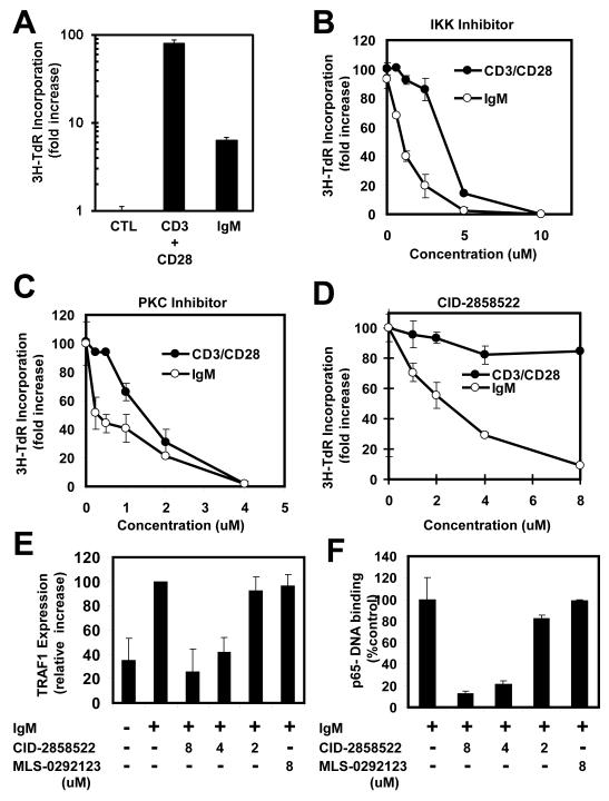
Dysregulation of NF-kappaB activity contributes to many autoimmune and inflammatory diseases. At least nine pathways for NF-kappaB activation have been identified, most of which converge on the IkappaB kinases (IKKs). Although IKKs represent logical targets for potential drug discovery, chemical inhibitors of IKKs suppress all known NF-kappaB activation pathways and thus lack the selectivity required for safe use. A unique NF-kappaB activation pathway is initiated by protein kinase C (PKC) that is stimulated by antigen receptors and many growth factor receptors. Using a cell-based high-throughput screening (HTS) assay and chemical biology strategy, we identified a 2-aminobenzimidazole compound, CID-2858522, which selectively inhibits the NF-kappaB pathway induced by PKC, operating downstream of PKC but upstream of IKKbeta, without inhibiting other NF-kappaB activation pathways. In human B cells stimulated through surface immunoglobulin, CID-2858522 inhibited NF-kappaB DNA-binding activity and expression of endogenous NF-kappaB-dependent target gene, TRAF1. Altogether, as a selective chemical inhibitor of the NF-kappaB pathway induced by PKC, CID-2858522 serves as a powerful research tool and may reveal new paths toward therapeutically useful NF-kappaB inhibitors.


Figures

Figure 1
Figure 1. Flow chart of screening and counter-screenings for selective inhibitors of antigen-receptor induced NF-κB pathway
A total of 114,889 compounds were screened. From a screen of 53,280 commercially available compounds (left column), 519 hits were obtained, of which 248 reconfirmed using the same primary screening assay. Next, the same HEK293-NF-κB reporter gene cell line was stimulated with TNF, leaving 46 compounds that failed to inhibit. Four of these compounds showed ctyotoxic activity, leaving 42 compounds, of which 11 showed activity when fresh stocks were ordered and tested. From these 11 active compounds, one hit, CID-2858522, inhibited PMA/Ionomycin-induced production of cytokine IL-8 by the HEK293 cell line, (an additional screen of compounds from a 61,609 library provided by the NIH using the same HTS assay resulted in no compounds that fulfilled the desired criteria (http://pubchem.ncbi.nlm.gov/assay/assay.cgi?aid-465&loc=ea_ras)). CID-2858522 was then characterized (right column). PDB was substituted for PMA to confirm suppression of an alternative PKC activator. Pathway selectivity was assessed using panels of cell lines, starting with HEK293 cells in which each of the remaining NF-κB activation pathways was stimulated (see Supplemental Figure 2 for details), showing inhibition only of PMA/Ionomycin-induced NF-κB reporter gene activity (panel 1). This was followed by a panel of secondary assays using various cell lines or primary cultured splenocytes (panel 2), measuring various end-points, which included NF-κB luciferase reporter gene activity (“Luc”), cytokine secretion, and 3H-Thymidine (3H-TdR), following stimulation with various inducers of specific upstream activators of NF-κB signaling, including agonists of TCRs (anti-CD3/CD28 for Jurkat, splenocytes), BCRs (anti-IgM for splenocytes), TLRs (LPS for THP.1 monocytes), TNFRs that signal through the “alternative” pathway (LTβR antibody for HeLa cells), and NLRs (γTriDAP for activating NOD1 and muramyl-dipeptide [MDP] for activating NOD2 in McF-7 breast cancer and THP.1 monocytes respectively). CID-285822 inhibited only anti-CD3/CD28-stimulated and anti-IgM-stimulated splenocytes (see Figures 3 and 4 for details). Various in vitro kinase assays were employed, showing no inhibition, followed by a kinome screen using competitive displacement of ATP.
Figure 2
Figure 2. CID-2858522 inhibits NF-κB activation and IL-8 production induced by PKC activators
(A) Structures of two hit compounds, CID-2858522 (left) and CID-2998237 (right) are shown; (B, C) 293-NF-κB-luc cells were pretreated for 2hrs with various concentration of either CID-2858522 (B) or CID-29982387 (C) and then stimulated with TNF (10 ng/ml) or PMA/Ionomycin (10 ng/ml; 5 ng/mL) for 16 h. Luciferase activity was measured and data were expressed as a percentage relative to control treatment with DMSO only (mean ± SD; n=3). (D) 293-NF-κB-luc cells were pretreated for 2 h with various concentrations of CID-2858522 or CID-2998237, then stimulated with PMA/Ionomycin for 16 h. IL-8 release into the medium was measured, expressing data as a percentage relative to control cultures treated with DMSO (mean ± SD; n=3). (E) 293-NF-κB-luc cells were pretreated with CID-2858522 and then stimulated with PDBu for 16 h, IL-8 production and NF-κB luciferase activity were measured as above. (F) 293-NF-κB-luc cells were pretreated for 2 h with various concentrations of CID-2858522 or a PKC inhibitor Bisindolylmaleimide I (1 uM) followed by PMA/Ionomycin (10 ng/ml each) for 2 h, then p65-DNA-binding activity was measured in nuclear extracts (10 ug protein) using an ELISA method, expressing data as fold-increase relative to unstimulated cells (mean ± SD; n=3).
Figure 3
Figure 3. CID-2858522 inhibits IL-2 production induced by anti-CD3/CD28 or PMA/Ionomycin in Jurkat cells
(A) Jurkat T cells were cultured with anti-CD3/anti-CD28/anti-mouse IgG (6 ug/ml each) or PMA/Ionomysin (10 ng/ml; 5 ng/ml) for 24 h, then IL-2 production in medium was measured (mean ± SD; n=3). (B-D) Jurkat T cells were pretreated for 2 h with IKK inhibitor, BMS-335541 (B), PKC inhibitor, Bisindolylmaleimide I (C), or CID-2858522 (D) and then stimulated with anti-CD3/anti-CD28/anti-mouse IgG (6 ug/ml each) or PMA/Ionomycin (10 ng/ml; 5 ng/ml) for 24 h. IL-2 production in medium, expressing data as a percentage of control (mean ± SD; n=3). (E) Jurkat cells were cultured with CID-2858522, PKC inhibitor, or IKK inhibitor for 24 h, then cell viability was determined based on ATP levels, expressing data as a percentage relative to control cells cultured with DMSO only (mean ± SD; n=3).
Figure 4
Figure 4. CID-2858522 inhibits anti-IgM-induced NF-κB activation and proliferation of β-lymphocytes
(A) Isolated mouse primary splenocytes were cultured with anti-CD3/anti-CD28 (0.3 ug/ml each) or anti-IgM (3 ug/ml) for 48 h, then 1 uCi 3H-Thymidine was added for 12 h and incorporation into DNA was measured, expressing data as fold increase above unstimulated cells (mean ± SD; n=3). (B-D) Primary splenocytes were pretreated for 2 h with IKK inhibitor, BMS-335541 (B), PKC inhibitor, Bisindolylmaleimide I (C) or CID-2858522 (D), then stimulated with anti-CD3/anti-CD28 (0.3 ug/ml) or anti-IgM (3 ug/ml) for 48 h. 1 uCi 3H-Thymidine was added for 12 h and incorporation into DNA was measured, expressing data as percent inhibition relative to control cells treated with DMSO (mean ± SD; n=3). (E) Human CLL B cell samples (n=3) were cultured for 12 hrs with compound CID-2858522 or inactive analog MLS-0292123, then stimulated with biotin anti-IgM (10 ug/mL) for 24 hrs. Levels of TRAF1 and β-actin were assessed by immunoblotting, quantified by densitometry and TRAF1 results reported relative to control cells, after normalization for β-actin (mean ± SD). (F) Human CLL B-cells were treated with various concentrations of CID-285822 or its inactive analog, MLS-0292123, for 12 h followed by biotin-anti-IgM (10 ug/ml) and avidin (10 ug/ml) for 2 h. Then nuclear extracts were prepared and p65-DNA-binding activity was measured (mean ± SD; n=3).
Figure 5
Figure 5. CID-285852 inhibits IKKβ phosphorylation induced by PKC activators
(A) HEK293 cells were cultured in 0.5% FBS medium for 24 h and then treated with CID-2858522 (4 uM) or PKC inhibitor, Bisindolylmaleimide I (1 uM) for 2 hrs followed by PMA/Ionomycin (10 ng/ml; 5 ng/ml) for 2 h. Cell lysates were subjected to immunoprecipitation using anti-CARMA1 antibody and analyzed by immunoblotting with anti-phospho-CARMA1 antibody or anti-CARMA1 antibody. (B-E) HEK293 cells were transfected with plasmids encoding myc-CARMA1 in combination with plasmids encoding various other proteins including Caspase-8 (cys-87ala) (B), HA-IKK-γ (C), HA-TAK1 (D), and TRAF6 (E). After 36 h. cells were cultured in 0.5% FBS medium for 12 h and then treated with CID-2858522 (4 uM) or Bisindolylmaleimide I (1 uM) for 2 hrs, followed by PMA/Ionomycin treatment (10 ng/ml; 5 ng/ml) for 2 hrs. Cell lysates were immunoprecipated using anti-myc antibody and analyzed by immunoblotting using anti-Caspase8 (B), anti-HA (C,D), or anti-TRAF6 (E) antibodies. (F) HEK293 cells were treated with CID-2858522 (4 uM) or PKC inhibitor (1 uM) followed by PMA/Ionomycin (10 ng/ml; 5 ng/ml) treatment for 2 hrs. Cell lysates were normalized for protein content and analyzed by immunoblotting using anti-FLIP and anti-alpha-tubulin antibodies. (G) HEK293 cells were cultured in 0.5% FBS medium for 24 hrs, then treated with CID-2858522 (4 uM) or its inactive analog, MLS-0292123, (4 uM), or PKC inhibitor (1 uM), followed by treatment for 5 min with PMA/Ionomycin (10 ng/ml) or TNF (10 ng/ml). Cell lysates were immunoprecipitated using anti-IKK-β and analyzed by immunoblotting using anti-phospho-IKK-β antibody or anti-IKK-β antibody (as loading control). Approximate molecular weights of all proteins are indicated in kiloDaltons.

Similar articles

Cited by

References

    1. Tracey KJ, Cerami A. Tumor necrosis factor, other cytokines and disease. Annu Rev Cell Biol. 1993;9:317–43. - PubMed
    1. Karin M, Delhase M. The I kappa B kinase (IKK) and NF-kappa B: key elements of proinflammatory signalling. Semin Immunol. 2000;12(1):85–98. - PubMed
    1. West AP, Koblansky AA, Ghosh S. Recognition and signaling by toll-like receptors. Annu Rev Cell Dev Biol. 2006;22:409–37. - PubMed
    1. Fujita T. Virology. Sensing viral RNA amid your own. Science. 2006;314(5801):935–6. - PubMed
    1. Janssens S, Tschopp J. Signals from within: the DNA-damage-induced NF-kappaB response. Cell Death Differ. 2006;13(5):773–84. - PubMed

Publication types

MeSH terms