. 2010 Feb;124(1):55-68.
doi: 10.1037/a0018320.

Lesions of the rat perirhinal cortex spare the acquisition of a complex configural visual discrimination yet impair object recognition

Affiliations
Free PMC article

Lesions of the rat perirhinal cortex spare the acquisition of a complex configural visual discrimination yet impair object recognition

John P Aggleton et al. Behav Neurosci. 2010 Feb.
Free PMC article

Abstract

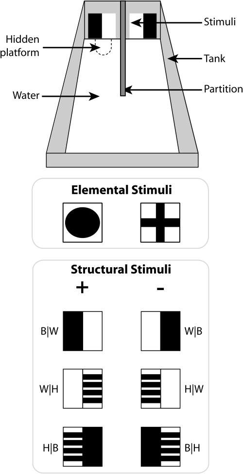
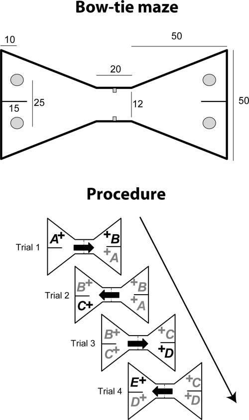
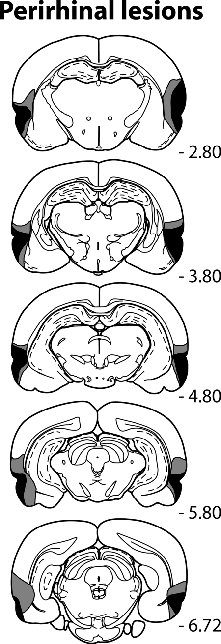
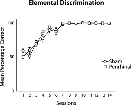
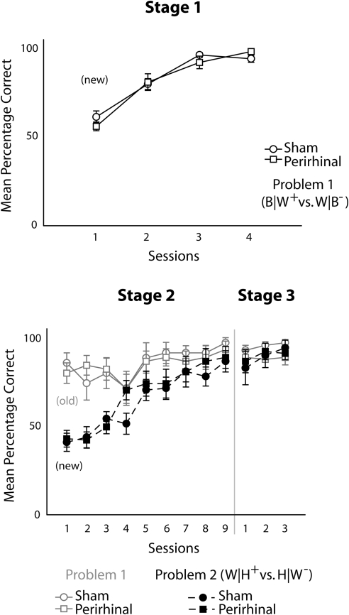
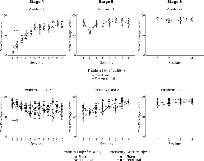
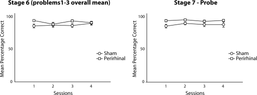
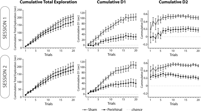
Rats with perirhinal cortex lesions were sequentially trained in a rectangular water tank on a series of 3 visual discriminations, each between mirror-imaged stimuli. When these same discriminations were tested concurrently, the rats were forced to use a configural strategy to solve the problems effectively. There was no evidence that lesions of the perirhinal cortex disrupted the ability to learn the concurrent configural discrimination task, which required the rats to learn the precise combination of stimulus identity with stimulus placement ("structural" learning). The same rats with perirhinal cortex lesions were also unimpaired on a test of spatial working memory (reinforced T maze alternation), although they were markedly impaired on a new test of spontaneous object recognition. For the recognition test, rats received multiple trials within a single session in which on every trial, they were allowed to explore 2 objects, 1 familiar, the other novel. On the basis of their differential exploration times, rats with perirhinal cortex lesions showed very poor discrimination of the novel objects, thereby confirming the effectiveness of the surgery. The discovery that bilateral lesions of the perirhinal cortex can leave configural (structural) learning seemingly unaffected points to a need to refine those models of perirhinal cortex function that emphasize its role in representing conjunctions of stimulus features.


Figures

Figure 1
Figure 1. Upper: Water tank apparatus used for testing the elemental and structural discriminations. Rats were placed in the water facing the near wall; discriminative stimuli were at the far end of the tank. An opaque partition separated the reinforced pattern (S+) from the nonreinforced pattern (S–). (The stimuli depicted are the first pair from the structural discrimination.) A submerged platform was always present underneath the reinforced pattern. Middle: The two stimuli used for the elemental discrimination, circle versus cross. The identity of the S+ stimulus was counterbalanced across the rats. Lower: The compound stimuli used for the structural discrimination task (Stages 1–6). Stimuli were formed from the elements black (B), white (W), and horizontal (H) presented in pairs with specific spatial relationships, for example, B|W, W|H, H|B. Reinforced stimuli are depicted in the left column (S+), and nonreinforced stimuli (S–) are depicted in the right column. Stimuli in the top row were used for Stage 1. Stimuli in the top two rows were used for Stages 2 and 3 of the structural discrimination, and all three rows of patterns were used in Stages 4–6. When all three discriminations were presented concurrently, every element was presented an equal number of times on the left or right of a compound stimulus as an S+ or S–. Thus, to solve the task the rat needed to learn the left and right positions of each specific pair of elements.
Figure 2
Figure 2. Object recognition memory. Upper: Shape and dimensions (in cm) of the bow-tie maze when viewed from above. Food wells are shown in gray. Lower: Illustration of the general test procedure showing the presentation order of the objects. All objects are rewarded (+). The arrows show the direction of the rats’ movements. Letters in black print represent novel objects; gray letters represent familiar objects.
Figure 3
Figure 3. Coronal sections depicting the extent of cell loss in the animals with the smallest (dark gray) and largest (light gray) perirhinal cortex lesions. The numbers refer to the approximate distance (in mm) of the section caudal to bregma. The sections are modified from The Rat Brain in Stereotaxic CoordinatesG. Paxinos & C. Watson, 1997
Figure 4
Figure 4. Elemental discrimination (circle vs. cross). Mean performance of the perirhinal lesion (PRh, square) and sham control (Sham, circle) groups. The vertical bars show the standard error of the mean (although when small, they are obscured by the symbols).
Figure 5
Figure 5. Structural discrimination learning, Stages 1–3. Mean performance of the perirhinal lesion (PRh, square) and sham control (Sham, circle) groups. The upper graph shows performance on the first discrimination (B|W vs. W|B). The lower graphs (Stages 2 and 3) show performance on the second (new) discrimination (Task 2, W|H vs. H|W) while testing on the previous discrimination (B|W vs. W|B) continued (“old,” light gray). The vertical bars show the standard error of the mean (although when small, they are obscured by the symbols). B = black; H = horizontal; W = white.
Figure 6
Figure 6. Structural discrimination learning, Stages 4–6. Mean performance of the perirhinal lesion (PRh, square) and sham control (Sham, circle) groups. The upper graphs show performance on the third (new) discrimination (H|B vs. B|H). The lower graphs show concurrent performance on the previous two structural discriminations (Tasks 1 and 2). In Stage 6, all three discriminations were equally intermingled within a session so that the rats had to solve the full configural task. The vertical bars show the standard error of the mean (although when small, they are obscured by the symbols). B = black; H = horizontal; W = white.
Figure 7
Figure 7. Structural discrimination learning. Left: Mean performance of the perirhinal lesion (PRh, square) and sham control (Sham, circle) groups on Stage 6 across all three concurrent, structural discriminations. Right: Stage 7 (probe). Mean performance when the compound stimuli were re-paired to give novel combinations of S+ with S– stimuli. Performance of both groups remained above chance. The vertical bars show the standard error of the mean (although when small, they are obscured by the symbols).
Figure 8
Figure 8. Object recognition. Six graphs depicting the mean performance from Session 1 (upper row) and Session 2 (lower row). Session 1 (upper): left, cumulative total exploration times for all objects; middle, cumulative D1 score; right, cumulative D2 score. Session 2 (lower): left, cumulative total exploration times for all objects; middle, cumulative D1 score; right, cumulative D2 score. Black symbols show the performance of the perirhinal lesion group, white symbols show the performance of the Sham control group. The vertical bars show the standard error of the mean (although when small, they are obscured by the symbols).

References

    1. Aggleton J. P. (in press). Understanding retrosplenial amnesia: Insights from animal studies. Neuropsychologia. - PubMed
    1. Aggleton J. P., Hunt P. R., & Rawlins J. N. P. (1986). The effects of hippocampal lesions upon spatial and nonspatial tests of working memory. Behavioural Brain Research, 19, 133–146. - PubMed
    1. Aggleton J. P., Keen S., Warburton E. C., & Bussey T. J. (1997). Extensive cytotoxic lesions of the rhinal cortices impair recognition but spare spatial alternation in the rat. Brain Research Bulletin, 43, 279–287. - PubMed
    1. Aggleton J. P., Kyd R., & Bilkey D. K. (2004). When is the perirhinal cortex necessary for the performance of spatial memory tasks? Neuroscience & Biobehavioral Reviews, 28, 611–624. - PubMed
    1. Aggleton J. P., Poirier G. L., Aggleton H. S., Vann S. D., & Pearce J. M. (2009). Lesions of the fornix and anterior thalamic nuclei dissociate different aspects of hippocampal-dependent spatial learning: Implications for the neural basis of scene learning. Behavioral Neuroscience, 123, 504–519. - PubMed

Publication types