

Liver sinusoidal endothelial fenestrations in caveolin-1 knockout mice
- PMID: 20141598
- PMCID: PMC4309280
- DOI: 10.1111/j.1549-8719.2009.00004.x
Liver sinusoidal endothelial fenestrations in caveolin-1 knockout mice
Abstract
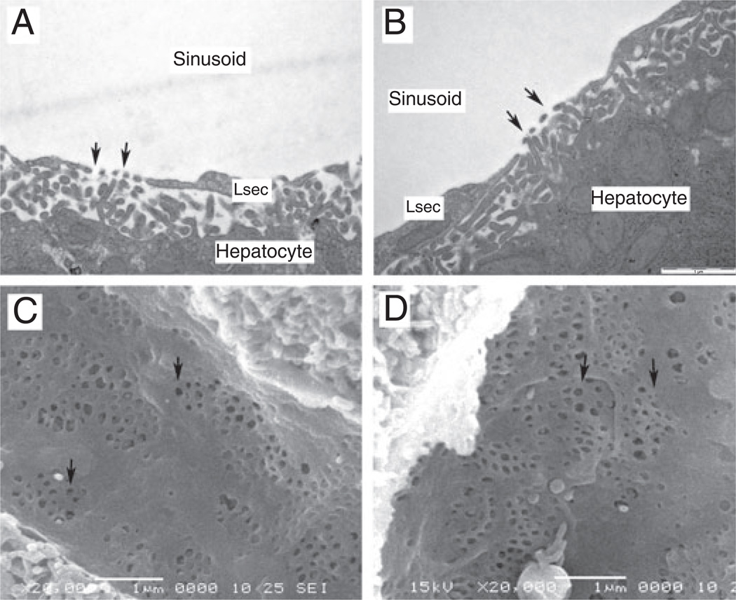
Objective: Fenestrations are pores in the liver sinusoidal endothelium that facilitate the transfer of particulate substrates between the sinusoidal lumen and hepatocytes. Fenestrations express caveolin-1 and have structural similarities to caveolae, therefore might be a form of caveolae and caveolin-1 may be integral to fenestration structure and function. Therefore, fenestrations were studied in the livers of caveolin-1 knockout mice.
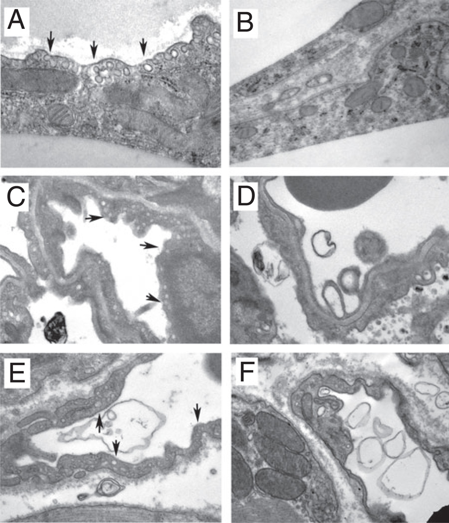
Methods: Scanning, transmission and immunogold electron microscopic techniques were used to study the liver sinusoidal endothelium and other tissues in caveolin-1 knockout and wild-type mice.
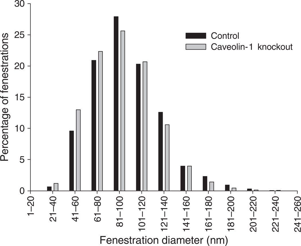
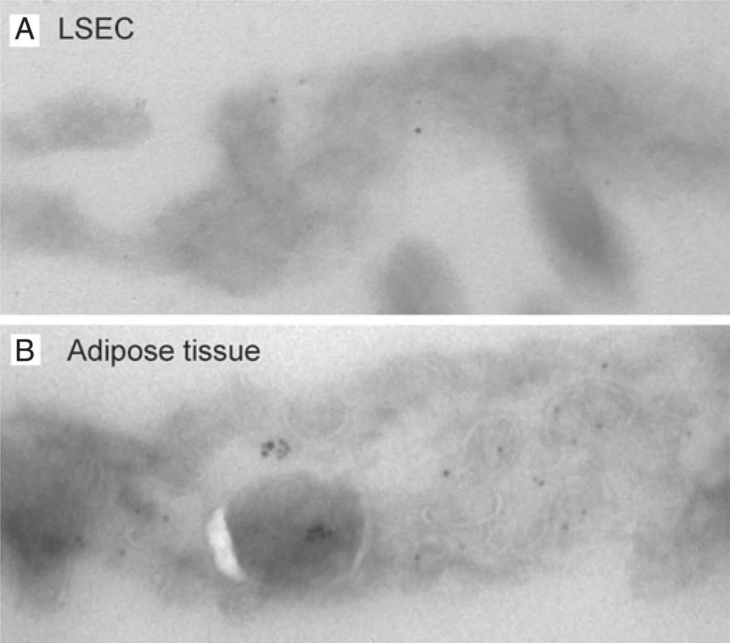
Results: Comparison of fenestrations in wild-type and knockout mice did not reveal any differences on either scanning or transmission electron microscopy. The diameter of the fenestrations was not significantly different (74 +/- 13 nm knockout mice vs 78 +/- 12 nm wild-type mice) nor was the fenestration porosity (6.5 +/- 2.1 knockout vs 7.3 +/- 2.4% wild-type mice). In contrast, adipocytes and blood vessels in other tissues lacked caveolae in the knockout mice. Caveolin-1 immunogold of livers of wild-type mice indicated sparse expression in sinusoidal endothelial cells.
Conclusions: The normal structure of fenestrations in the liver sinusoidal endothelium is not dependent upon caveolin-1 and fenestrations are not a form of caveolae.
Figures
Similar articles
-
Distribution and localization of caveolin-1 in sinusoidal cells in rat liver.Med Electron Microsc. 2003 Mar;36(1):33-40. doi: 10.1007/s007950300004. Med Electron Microsc. 2003. PMID: 12658349
-
Vascular endothelial growth factor increases fenestral permeability in hepatic sinusoidal endothelial cells.Liver Int. 2003 Dec;23(6):467-75. doi: 10.1111/j.1478-3231.2003.00880.x. Liver Int. 2003. PMID: 14986821
-
The effect of feeding and fasting on fenestrations in the liver sinusoidal endothelial cell.Pathology. 2010 Apr;42(3):255-8. doi: 10.3109/00313021003636469. Pathology. 2010. PMID: 20350219
-
Regulatory mechanisms of hepatic microcirculation.Clin Hemorheol Microcirc. 2003;29(3-4):167-82. Clin Hemorheol Microcirc. 2003. PMID: 14724338 Review.
-
Caveolae and endothelial dysfunction: filling the caves in cardiovascular disease.Eur J Pharmacol. 2008 May 13;585(2-3):256-60. doi: 10.1016/j.ejphar.2008.02.086. Epub 2008 Mar 15. Eur J Pharmacol. 2008. PMID: 18423600 Review.
Cited by
-
Cavin1 Deficiency Causes Disorder of Hepatic Glycogen Metabolism and Neonatal Death by Impacting Fenestrations in Liver Sinusoidal Endothelial Cells.Adv Sci (Weinh). 2020 Aug 21;7(19):2000963. doi: 10.1002/advs.202000963. eCollection 2020 Oct. Adv Sci (Weinh). 2020. PMID: 33042738 Free PMC article.
-
Scanning and transmission electron microscopy of the cells forming the hepatic sinusoidal wall of rat in acetaminophen and Escherichia coli endotoxin-induced hepatotoxicity.J Microsc Ultrastruct. 2017 Jan-Mar;5(1):21-27. doi: 10.1016/j.jmau.2016.04.003. Epub 2016 May 7. J Microsc Ultrastruct. 2017. PMID: 30023233 Free PMC article.
-
Three-dimensional structured illumination microscopy of liver sinusoidal endothelial cell fenestrations.J Struct Biol. 2010 Sep;171(3):382-8. doi: 10.1016/j.jsb.2010.06.001. Epub 2010 Jun 4. J Struct Biol. 2010. PMID: 20570732 Free PMC article.
-
Autophagic degradation of caveolin-1 promotes liver sinusoidal endothelial cells defenestration.Cell Death Dis. 2018 May 1;9(5):576. doi: 10.1038/s41419-018-0567-0. Cell Death Dis. 2018. PMID: 29760379 Free PMC article.
-
Caveolin 1-related autophagy initiated by aldosterone-induced oxidation promotes liver sinusoidal endothelial cells defenestration.Redox Biol. 2017 Oct;13:508-521. doi: 10.1016/j.redox.2017.07.011. Epub 2017 Jul 13. Redox Biol. 2017. PMID: 28734243 Free PMC article.
References
-
- Arias IM. The biology of hepatic endothelial cell fenestrae. Prog Liver Dis. 1990;9:11–26. - PubMed
-
- Braet F, De Zanger R, Baekeland M, Crabbe E, Van Der Smissen P, Wisse E. Structure and dynamics of the fenestrae-associated cytoskeleton of rat liver sinusoidal endothelial cells. Hepatology. 1995;21:180–189. - PubMed
-
- Cogger VC, Le Couteur DG. Fenestrations in the liver sinusoidal endothelial cell. In: Arias I, Wolkoff A, Boyer J, Shafritz D, Fausto N, Alter H, et al., editors. The Liver: Biology and Pathobiology. 5th edn. Hokoben, NJ: John Wiley & Sons, Ltd; 2009. pp. 387–404.
Publication types
MeSH terms
Substances
Grants and funding
LinkOut - more resources
Full Text Sources
Other Literature Sources
Molecular Biology Databases
