. 2009 Aug;13(8B):1857-65.
doi: 10.1111/j.1582-4934.2009.00644.x.

Calcium and ER stress mediate hepatic apoptosis after burn injury

Affiliations

Calcium and ER stress mediate hepatic apoptosis after burn injury

Marc G Jeschke et al. J Cell Mol Med. 2009 Aug.

Abstract

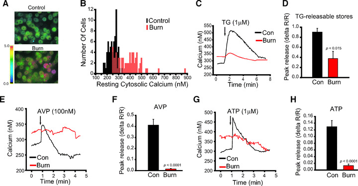
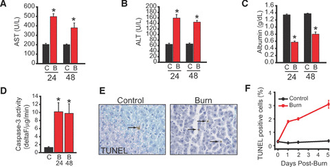
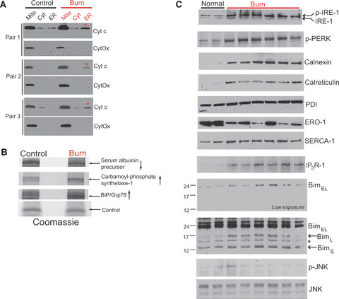
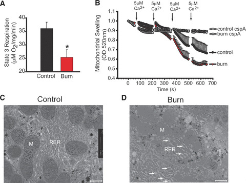
A hallmark of the disease state following severe burn injury is decreased liver function, which results in gross metabolic derangements that compromise patient survival. The underlying mechanisms leading to hepatocyte dysfunction after burn are essentially unknown. The aim of the present study was to determine the underlying mechanisms leading to hepatocyte dysfunction and apoptosis after burn. Rats were randomized to either control (no burn) or burn (60% total body surface area burn) and sacrificed at various time-points. Liver was either perfused to isolate primary rat hepatocytes, which were used for in vitro calcium imaging, or liver was harvested and processed for immunohistology, transmission electron microscopy, mitochondrial isolation, mass spectroscopy or Western blotting to determine the hepatic response to burn injury in vivo. We found that thermal injury leads to severely depleted endoplasmic reticulum (ER) calcium stores and consequent elevated cytosolic calcium concentrations in primary hepatocytes in vitro. Burn-induced ER calcium depletion caused depressed hepatocyte responsiveness to signalling molecules that regulate hepatic homeostasis, such as vasopressin and the purinergic agonist ATP. In vivo, thermal injury resulted in activation of the ER stress response and major alterations in mitochondrial structure and function - effects which may be mediated by increased calcium release by inositol 1,4,5-trisphosphate receptors. Our results reveal that thermal injury leads to dramatic hepatic disturbances in calcium homeostasis and resultant ER stress leading to mitochondrial abnormalities contributing to hepatic dysfunction and apoptosis after burn injury.

Keywords: ER stress; apoptosis; calcium; liver; thermal injury; unfolded protein response.


Figures

Figure 2
Figure 2
Burn injury results in significant alterations of hepatocyte calcium homeostasis. (A) Representative fura‐2 ratio images of cytosolic calcium in isolated hepatocytes from a control and an animal 24 hrs after burn injury. Ratio values are pseudo‐coloured as per the scale bar. (B) Histogram of resting cytosolic calcium in control (black bars) and burned (red bars) hepatocytes (n= 70 for each condition). (C) Single cell imaging of depletion of endoplasmic reticulum calcium stores with thapsigargin (TG) in control hepatocytes (black) and hepatocytes isolated from burned animals (red). Each trace is an average of 20 hepatocytes from a single experiment. (D) Quantified TG‐releasable stores in hepatocytes isolated from control (black) and burned (red) animals pooled from three separate experiments. Peak release is quantified as the change in ratio (ΔR) divided by the initial ratio (R). (E) Single cell imaging of hepatocyte responses to Arg‐vasopressin (AVP) in hepatocytes isolated from control (black) and burned (red) animals. (F) Quantified responses to vasopressin in hepatocytes isolated from control (black) and burned (red) animals. (G) Single cell imaging of hepatocyte responses to extracellular ATP in hepatocytes isolated from control (black) and burned (red) animals. (H) Quantified responses to ATP in hepatocytes isolated from control (black) and burned (red) animals.
Figure 1
Figure 1
Burn injury causes hepatic damage, dysfunction and hepatocyte apoptosis. Hepatic dysfunction of the rat burn model mimics the post‐burn human disease state. (A) Serum aspartate aminotransferase (AST) 24 and 48 hrs after burn injury (each time‐point represent eight animals per group). (B) Serum alanine aminotransferase (ALT) 24 and 48 hrs after burn injury (each time‐point represent eight animals per group). (C) Serum albumin 24 and 48 hrs after burn injury (each time‐point represent eight animals per group). (D) Caspase‐3 activity in liver cytosolic fractions was determined by fluorometric Asp‐Glu‐Val‐Asp cleavage assays, as described previously [22]. The data represent the average of three separate experiments. Each experiment was performed in triplicate. (E) TUNEL staining of a liver section before (control) and 24 hrs after burn injury (burn). Arrow indicates TUNEL positive cells (F) Quantified TUNEL‐positive cells 24, 48 and 120 hrs after burn injury (each time‐point represent eight animals per group). Time after injury in hours is indicated. C, control; B, burn. Data in this and subsequent figures are the mean ± S.E.M. *P < 0.05.
Figure 3
Figure 3
Thermal injury induces hepatic endoplasmic reticulum (ER) stress response in vivo. (A) Cytochrome c and cytochrome c oxidase (CytOx) distribution in mitochondrial (mito), cytosolic (cyt) and ER fractions from three pairs of control and burned animals 24 hrs after injury. Cytochrome c appearance in the ER fraction is indicated by a red asterisk. (B) Proteins which display prominent alterations 24 hrs after burn injury. Bands are detected by Coomassie staining. Band identities were determined by trypsin digestion and MALDI‐TOF. (C) Western blots of ER stress proteins and ER calcium handling/chaperone proteins and/or their phosphoprotein levels in liver samples isolated from two control and six burned animals 24 hrs after burn injury. All Western blots are from the same animals. *Non‐specific band.
Figure 4
Figure 4
Thermal injury induces hepatic mitochondrial dysfunction in vivo. (A) State 3 respiration in isolated mitochondria from animals 48 hrs after burn injury (red) or in control animals (black). (B) Calcium‐induced swelling in mitochondria isolated 48 hrs after burn injury. Isolated mitochondria were challenged in bulk solution with successive additions of 5 μM calcium chloride as indicated. Mitochondrial swelling was monitored by measuring the optical density at 520 nm (see Experimental Procedures). Decreases in optical density indicate mitochondrial swelling. Cyclosporine A (cspA; 5 μM), a blocker of permeability transition pore (PTP) opening, was used to confirm that calcium‐induced swelling was due to PTP opening. Data represent the pooled responses from three animals in each treatment group. Note the earlier onset and more rapid and complete swelling of mitochondria isolated from burned animals. (C) Transmission electron microscope images from livers of control animal. Note prominent mitochondrial cristae and the surrounding rough ER. M, mitochondria; RER, rough endoplasmic reticulum. (D) Transmission electron microscope images of liver sections from an animal 48 hrs after burn injury. Note prominent loss of mitochondrial electron density with lack of cristae. Focal dilation of rough ER is clearly apparent and indicated by arrows. Both micrographs were taken at the same magnification. Scale bars are 500 nm.

Similar articles

Cited by

References

    1. Brigham PA, McLoughlin E. Burn incidence and medical care use in the United States: estimates, trends and data sources. J Burn Care Rehabil. 1996; 17: 95–107. - PubMed
    1. World Health Organization (WHO) . A graphical overview of the global burden of injuries. The injury chart book. Volume 29. Geneva : WHO ; 2002.
    1. Herndon DN, Tompkins RG. Support of the metabolic response to burn injury. Lancet. 2004; 363: 1895–902. - PubMed
    1. Herndon DN, Hart DW, Wolf SE, et al . Reversal of catabolism by beta‐blockade after severe burns. N Engl J Med. 2001; 345: 1223–9. - PubMed
    1. van den Berge G, Wouters P, Weekers F, et al . Intensive insulin therapy in critically ill patients. New Engl J Med. 2001; 345: 1359–67. - PubMed

Publication types