

Pseudomonas aeruginosa cystic fibrosis isolates of similar RAPD genotype exhibit diversity in biofilm forming ability in vitro
- PMID: 20141637
- PMCID: PMC2841157
- DOI: 10.1186/1471-2180-10-38
Pseudomonas aeruginosa cystic fibrosis isolates of similar RAPD genotype exhibit diversity in biofilm forming ability in vitro
Abstract
Background: Pseudomonas aeruginosa is considered to grow in a biofilm in cystic fibrosis (CF) chronic lung infections. Bacterial cell motility is one of the main factors that have been connected with P. aeruginosa adherence to both biotic and abiotic surfaces. In this investigation, we employed molecular and microscopic methods to determine the presence or absence of motility in P. aeruginosa CF isolates, and statistically correlated this with their biofilm forming ability in vitro.
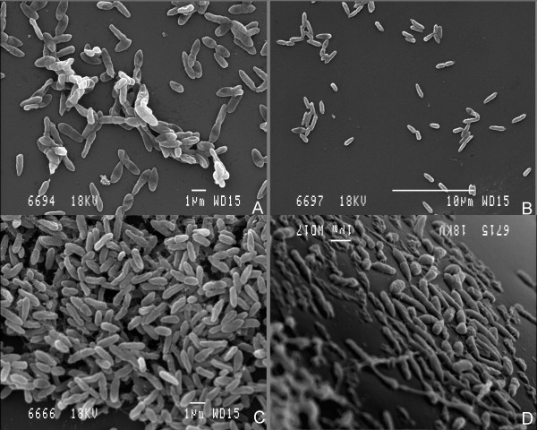
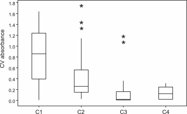
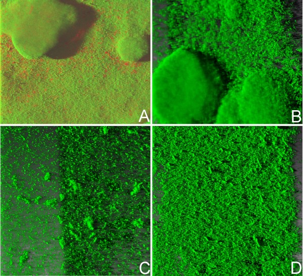
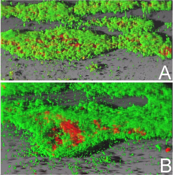
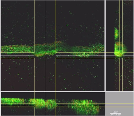
Results: Our investigations revealed a wide diversity in the production, architecture and control of biofilm formation. Of 96 isolates, 49% possessed swimming motility, 27% twitching and 52% swarming motility, while 47% were non-motile. Microtitre plate assays for biofilm formation showed a range of biofilm formation ability from biofilm deficient phenotypes to those that formed very thick biofilms. A comparison of the motility and adherence properties of individual strains demonstrated that the presence of swimming and twitching motility positively affected biofilm biomass. Crucially, however, motility was not an absolute requirement for biofilm formation, as 30 non-motile isolates actually formed thick biofilms, and three motile isolates that had both flagella and type IV pili attached only weakly. In addition, CLSM analysis showed that biofilm-forming strains of P. aeruginosa were in fact capable of entrapping non-biofilm forming strains, such that these 'non-biofilm forming' cells could be observed as part of the mature biofilm architecture.
Conclusions: Clinical isolates that do not produce biofilms in the laboratory must have the ability to survive in the patient lung. We propose that a synergy exists between isolates in vivo, which allows "non biofilm-forming" isolates to be incorporated into the biofilm. Therefore, there is the potential for strains that are apparently non-biofilm forming in vitro to participate in biofilm-mediated pathogenesis in the CF lung.
Figures
Similar articles
-
[Investigation of biofilm formation and relationship with genotype and antibiotic susceptibility of Pseudomonas aeruginosa strains isolated from patients with cystic fibrosis].Mikrobiyol Bul. 2009 Oct;43(4):563-73. Mikrobiyol Bul. 2009. PMID: 20084909 Turkish.
-
Cross-sectional analysis of clinical and environmental isolates of Pseudomonas aeruginosa: biofilm formation, virulence, and genome diversity.Infect Immun. 2004 Jan;72(1):133-44. doi: 10.1128/IAI.72.1.133-144.2004. Infect Immun. 2004. PMID: 14688090 Free PMC article.
-
Twitching motility activity, biofilm formation, and genetic typing for clinical isolates of Pseudomonas aeruginosa by random amplified DNA PCR.Acta Microbiol Immunol Hung. 2013 Sep;60(3):313-28. doi: 10.1556/AMicr.60.2013.3.7. Acta Microbiol Immunol Hung. 2013. PMID: 24060555
-
Pseudomonas aeruginosa chromosomal beta-lactamase in patients with cystic fibrosis and chronic lung infection. Mechanism of antibiotic resistance and target of the humoral immune response.APMIS Suppl. 2003;(116):1-47. APMIS Suppl. 2003. PMID: 14692154 Review.
-
Diagnosis of biofilm infections in cystic fibrosis patients.APMIS. 2017 Apr;125(4):339-343. doi: 10.1111/apm.12689. APMIS. 2017. PMID: 28407432 Review.
Cited by
-
Role of the flagellar hook in the structural development and antibiotic tolerance of Pseudomonas aeruginosa biofilms.ISME J. 2022 Apr;16(4):1176-1186. doi: 10.1038/s41396-021-01157-9. Epub 2021 Dec 8. ISME J. 2022. PMID: 34880458 Free PMC article.
-
Motility-Independent Formation of Antibiotic-Tolerant Pseudomonas aeruginosa Aggregates.Appl Environ Microbiol. 2019 Jul 1;85(14):e00844-19. doi: 10.1128/AEM.00844-19. Print 2019 Jul 15. Appl Environ Microbiol. 2019. PMID: 31076438 Free PMC article.
-
Genotypic and phenotypic analyses of a Pseudomonas aeruginosa chronic bronchiectasis isolate reveal differences from cystic fibrosis and laboratory strains.BMC Genomics. 2015 Oct 30;16:883. doi: 10.1186/s12864-015-2069-0. BMC Genomics. 2015. PMID: 26519161 Free PMC article.
-
Pseudomonas aeruginosa isolates from dental unit waterlines can be divided in two distinct groups, including one displaying phenotypes similar to isolates from cystic fibrosis patients.Front Microbiol. 2015 Jan 21;5:802. doi: 10.3389/fmicb.2014.00802. eCollection 2014. Front Microbiol. 2015. PMID: 25653647 Free PMC article.
-
Microfluidic approaches to bacterial biofilm formation.Molecules. 2012 Aug 15;17(8):9818-34. doi: 10.3390/molecules17089818. Molecules. 2012. PMID: 22895027 Free PMC article. Review.
References
Publication types
MeSH terms
LinkOut - more resources
Full Text Sources
Medical
Molecular Biology Databases
