

Recognition and binding of a helix-loop-helix peptide to carbonic anhydrase occurs via partly folded intermediate structures
- PMID: 20141756
- PMCID: PMC2814212
- DOI: 10.1016/j.bpj.2009.10.038
Recognition and binding of a helix-loop-helix peptide to carbonic anhydrase occurs via partly folded intermediate structures
Abstract
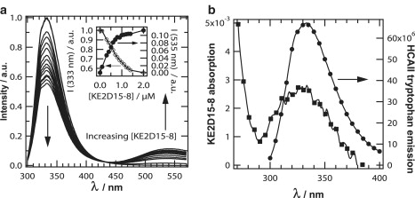
We have studied the association of a helix-loop-helix peptide scaffold carrying a benzenesulfonamide ligand to carbonic anhydrase using steady-state and time-resolved fluorescence spectroscopy. The helix-loop-helix peptide, developed for biosensing applications, is labeled with the fluorescent probe dansyl, which serves as a polarity-sensitive reporter of the binding event. Using maximum entropy analysis of the fluorescence lifetime of dansyl at 1:1 stoichiometry reveals three characteristic fluorescence lifetime groups, interpreted as differently interacting peptide/protein structures. We characterize these peptide/protein complexes as mostly bound but unfolded, bound and partly folded, and strongly bound and folded. Furthermore, analysis of the fluorescence anisotropy decay resulted in three different dansyl rotational correlation times, namely 0.18, 1.2, and 23 ns. Using the amplitudes of these times, we can correlate the lifetime groups with the corresponding fluorescence anisotropy component. The 23-ns rotational correlation time, which appears with the same amplitude as a 17-ns fluorescence lifetime, shows that the dansyl fluorophore follows the rotational diffusion of carbonic anhydrase when it is a part of the folded peptide/protein complex. A partly folded and partly hydrated interfacial structure is manifested in an 8-ns dansyl fluorescence lifetime and a 1.2-ns rotational correlation time. This structure, we believe, is similar to a molten-globule-like interfacial structure, which allows segmental movement and has a higher degree of solvent exposure of dansyl. Indirect excitation of dansyl on the helix-loop-helix peptide through Förster energy transfer from one or several tryptophans in the carbonic anhydrase shows that the helix-loop-helix scaffold binds to a tryptophan-rich domain of the carbonic anhydrase. We conclude that binding of the peptide to carbonic anhydrase involves a transition from a disordered to an ordered structure of the helix-loop-helix scaffold.
Copyright (c) 2010 Biophysical Society. Published by Elsevier Inc. All rights reserved.
Figures


Similar articles
-
A versatile polypeptide platform for integrated recognition and reporting: affinity arrays for protein-ligand interaction analysis.Chemistry. 2004 May 17;10(10):2375-85. doi: 10.1002/chem.200305391. Chemistry. 2004. PMID: 15146511
-
Designed, functionalized helix-loop-helix motifs that bind human carbonic anhydrase II: a new class of synthetic receptor molecules.J Am Chem Soc. 2004 Apr 14;126(14):4464-5. doi: 10.1021/ja038799c. J Am Chem Soc. 2004. PMID: 15070333
-
The slow folding reaction of barstar: the core tryptophan region attains tight packing before substantial secondary and tertiary structure formation and final compaction of the polypeptide chain.J Mol Biol. 2000 Sep 15;302(2):479-95. doi: 10.1006/jmbi.2000.4060. J Mol Biol. 2000. PMID: 10970747
-
Designed, folded polypeptide scaffolds that combine key biosensing events of recognition and reporting.J Org Chem. 2002 May 3;67(9):3120-3. doi: 10.1021/jo010954n. J Org Chem. 2002. PMID: 11975577 No abstract available.
-
The binding of human carbonic anhydrase II by functionalized folded polypeptide receptors.Chem Biol. 2005 Nov;12(11):1245-52. doi: 10.1016/j.chembiol.2005.08.018. Chem Biol. 2005. PMID: 16298304
Cited by
-
Cholesterol promotes the formation of dimers and oligomers of the receptor tyrosine kinase ROR1.bioRxiv [Preprint]. 2025 Jun 22:2025.06.19.660507. doi: 10.1101/2025.06.19.660507. bioRxiv. 2025. PMID: 40667148 Free PMC article. Preprint.
-
Difunctional Fluorescent Probes for Iron and Hydrogen Sulfide Detection Based on Diphenyl Derivative.J Fluoresc. 2024 May;34(3):1269-1278. doi: 10.1007/s10895-023-03374-1. Epub 2023 Aug 1. J Fluoresc. 2024. PMID: 37526873
-
Dynamic fluorescence depolarization: a powerful tool to explore protein folding on the ribosome.Methods. 2010 Sep;52(1):57-73. doi: 10.1016/j.ymeth.2010.06.001. Epub 2010 Jun 8. Methods. 2010. PMID: 20685617 Free PMC article. Review.
References
-
- Gavin A.-C., Aloy P., Superti-Furga G. Proteome survey reveals modularity of the yeast cell machinery. Nature. 2006;440:631–636. - PubMed
-
- Levy E.D., Pereira-Leal J.B. Evolution and dynamics of protein interactions and networks. Curr. Opin. Struct. Biol. 2008;18:349–357. - PubMed
-
- Pauling L. A theory of the structure and process of formation of antibodies. J. Am. Chem. Soc. 1940;62:2643–2657.
Publication types
MeSH terms
Substances
LinkOut - more resources
Full Text Sources
Miscellaneous
