Aldehyde dehydrogenase 1 A1-positive cell population is enriched in tumor-initiating cells and associated with progression of bladder cancer
- PMID: 20142235
- PMCID: PMC3544173
- DOI: 10.1158/1055-9965.EPI-09-0865
Aldehyde dehydrogenase 1 A1-positive cell population is enriched in tumor-initiating cells and associated with progression of bladder cancer
Abstract
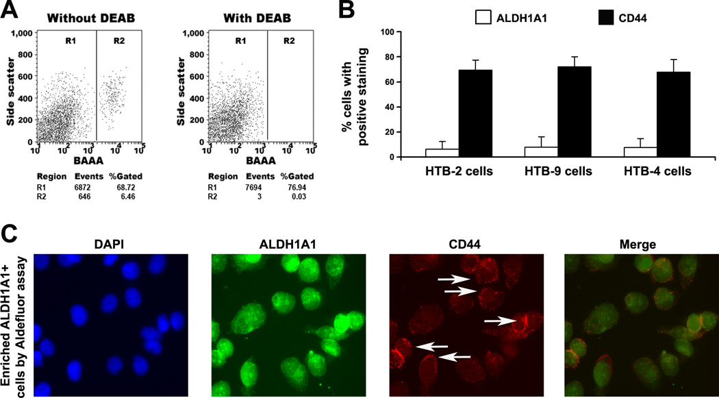
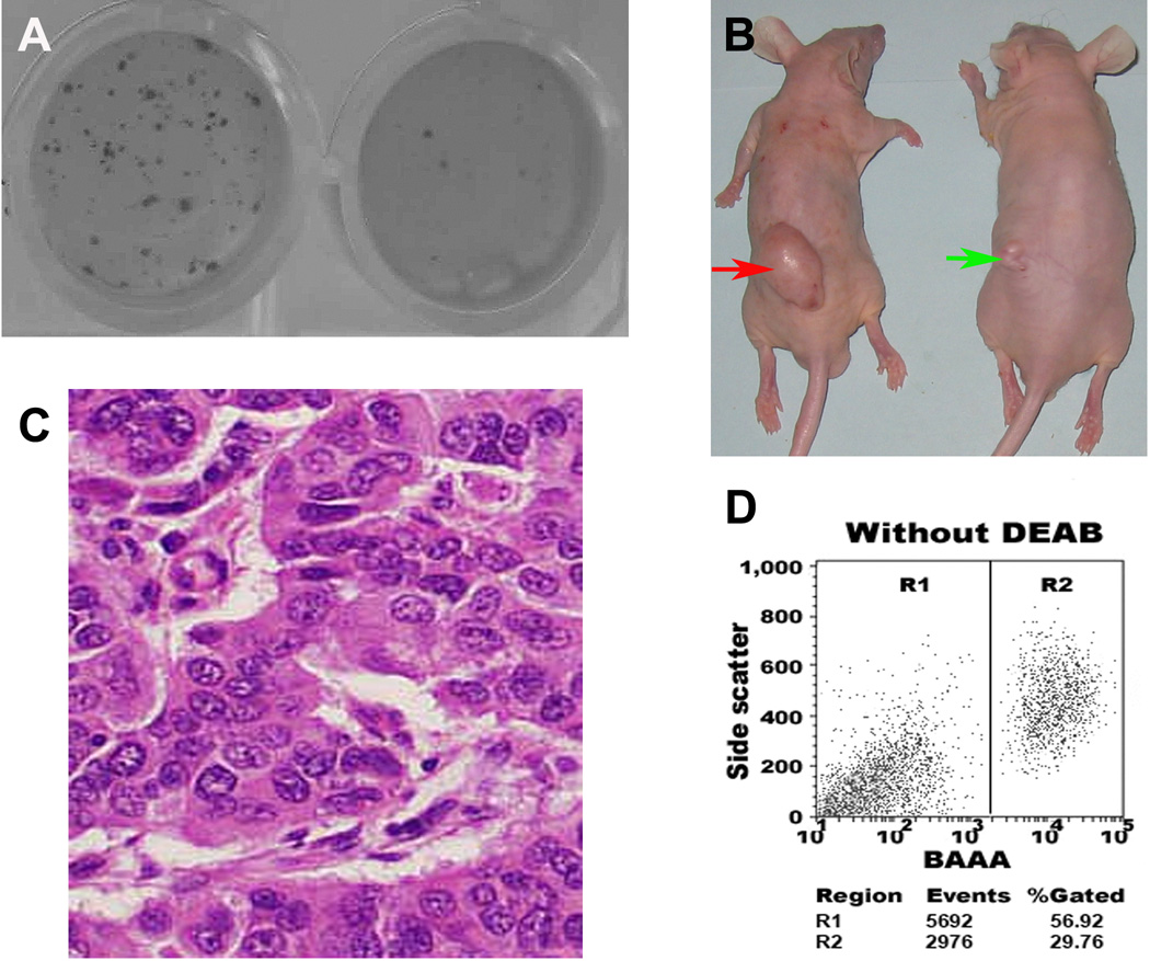
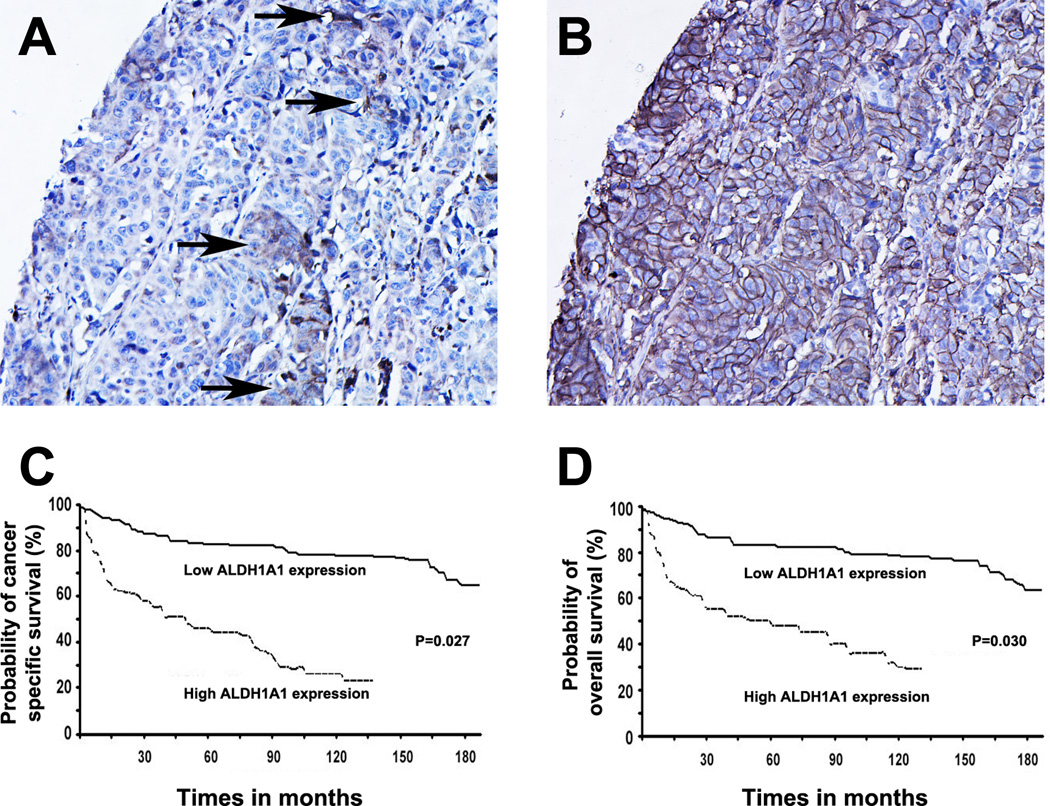
Aldehyde dehydrogenase 1 A1 (ALDH1A1) has recently been suggested as a marker for cancer stem or stem-like cancer cells of some human malignancies. The purpose of this study was to investigate the stem cell-related function and clinical significance of the ALDH1A1 in bladder urothelial cell carcinoma. Aldefluor assay was used to isolate ALDH1A1+ cells from bladder cancer cells. Stem cell characteristics of the ALDH1A1+ cells were then investigated by in vitro and in vivo approaches. Immunohistochemistry was done for evaluating ALDH1A1 expression on 22 normal bladder tissues and 216 bladder tumor specimens of different stage and grade. The ALDH1A1+ cancer cells displayed higher in vitro tumorigenicity compared with isogenic ALDH1A1- cells. The ALDH1A1+ cancer cells could generate xenograft tumors that resembled the histopathologic characteristics and heterogeneity of the parental cells. High ALDH1A1 expression was found in 26% (56 of 216) of human bladder tumor specimens and significantly related to advanced pathologic stage, high histologic grade, recurrence and progression, and metastasis of bladder urothelial cell carcinomas (all P < 0.05). Furthermore, ALDH1A1 expression was inversely associated with cancer-specific and overall survivals of the patients (P = 0.027 and 0.030, respectively). Therefore, ALDH1A1+ cell population could be enriched in tumor-initiating cells. ALDH1A1 may serve as a useful marker for monitoring the progression of bladder tumor and identifying bladder cancer patients with poor prognosis who might benefit from adjuvant and effective treatments.
Conflict of interest statement
Figures
Similar articles
-
ALDH1A1 is a marker for malignant prostate stem cells and predictor of prostate cancer patients' outcome.Lab Invest. 2010 Feb;90(2):234-44. doi: 10.1038/labinvest.2009.127. Epub 2009 Dec 14. Lab Invest. 2010. PMID: 20010854 Free PMC article.
-
ALDH1 in combination with CD44 as putative cancer stem cell markers are correlated with poor prognosis in urothelial carcinoma of the urinary bladder.Asian Pac J Cancer Prev. 2014;15(5):2013-20. doi: 10.7314/apjcp.2014.15.5.2013. Asian Pac J Cancer Prev. 2014. PMID: 24716927
-
Aldehyde dehydrogenase 1 is a tumor stem cell-associated marker in lung cancer.Mol Cancer Res. 2009 Mar;7(3):330-8. doi: 10.1158/1541-7786.MCR-08-0393. Epub 2009 Mar 10. Mol Cancer Res. 2009. PMID: 19276181 Free PMC article.
-
Normal and neoplastic urothelial stem cells: getting to the root of the problem.Nat Rev Urol. 2012 Oct;9(10):583-94. doi: 10.1038/nrurol.2012.142. Epub 2012 Aug 14. Nat Rev Urol. 2012. PMID: 22890301 Free PMC article. Review.
-
ALDH1A1 as a marker for metastasis initiating cells: A mechanistic insight.Exp Cell Res. 2024 Sep 1;442(1):114213. doi: 10.1016/j.yexcr.2024.114213. Epub 2024 Aug 22. Exp Cell Res. 2024. PMID: 39173941 Review.
Cited by
-
ALDEFLUOR activity, ALDH isoforms, and their clinical significance in cancers.J Enzyme Inhib Med Chem. 2023 Dec;38(1):2166035. doi: 10.1080/14756366.2023.2166035. J Enzyme Inhib Med Chem. 2023. PMID: 36651035 Free PMC article.
-
The Origin and Evolution of Bladder Cancer Stem Cells.Front Cell Dev Biol. 2022 Jul 12;10:950241. doi: 10.3389/fcell.2022.950241. eCollection 2022. Front Cell Dev Biol. 2022. PMID: 35903544 Free PMC article. Review.
-
Phenethyl Isothiocyanate Exposure Promotes Oxidative Stress and Suppresses Sp1 Transcription Factor in Cancer Stem Cells.Int J Mol Sci. 2019 Feb 27;20(5):1027. doi: 10.3390/ijms20051027. Int J Mol Sci. 2019. PMID: 30818757 Free PMC article.
-
Targeting of alpha-v integrins reduces malignancy of bladder carcinoma.PLoS One. 2014 Sep 23;9(9):e108464. doi: 10.1371/journal.pone.0108464. eCollection 2014. PLoS One. 2014. PMID: 25247809 Free PMC article.
-
Activation of D2 Dopamine Receptors in CD133+ve Cancer Stem Cells in Non-small Cell Lung Carcinoma Inhibits Proliferation, Clonogenic Ability, and Invasiveness of These Cells.J Biol Chem. 2017 Jan 13;292(2):435-445. doi: 10.1074/jbc.M116.748970. Epub 2016 Dec 5. J Biol Chem. 2017. PMID: 27920206 Free PMC article.
References
-
- Parkin DM, Bray F, Ferlay J, Pisani P. Estimating the world cancer burden: Globocan 2000. Int J Cancer. 2001;94:153–156. - PubMed
-
- Wu XR. Urothelial tumorigenesis: a tale of divergent pathways. Nat Rev Cancer. 2005;5:713–725. - PubMed
-
- Habuchi T, Marberger M, Droller MJ, et al. Prognostic markers for bladder cancer: International Consensus Panel on bladder tumor markers. Urology. 2005;66:64–74. - PubMed
-
- Malats N, Bustos A, Nascimento CM, et al. P53 as a prognosticmarker for bladder cancer: a meta-analysis and review. Lancet Oncol. 2005;6:678–686. - PubMed
-
- Jordan CT, Guzman ML, Noble M. Cancer stem cells. N Engl J Med. 2006;12:1253–1261. - PubMed
Publication types
MeSH terms
Substances
Grants and funding
LinkOut - more resources
Full Text Sources
Medical
Miscellaneous