Screen-film mammographic density and breast cancer risk: a comparison of the volumetric standard mammogram form and the interactive threshold measurement methods
- PMID: 20142240
- PMCID: PMC2875111
- DOI: 10.1158/1055-9965.EPI-09-1059
Screen-film mammographic density and breast cancer risk: a comparison of the volumetric standard mammogram form and the interactive threshold measurement methods
Abstract
Background: Mammographic density is a strong risk factor for breast cancer, usually measured by an area-based threshold method that dichotomizes the breast area on a mammogram into dense and nondense regions. Volumetric methods of breast density measurement, such as the fully automated standard mammogram form (SMF) method that estimates the volume of dense and total breast tissue, may provide a more accurate density measurement and improve risk prediction.
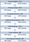
Methods: In 2000-2003, a case-control study was conducted of 367 newly confirmed breast cancer cases and 661 age-matched breast cancer-free controls who underwent screen-film mammography at several centers in Toronto, Canada. Conditional logistic regression was used to estimate odds ratios of breast cancer associated with categories of mammographic density, measured with both the threshold and the SMF (version 2.2beta) methods, adjusting for breast cancer risk factors.
Results: Median percent density was higher in cases than in controls for the threshold method (31% versus 27%) but not for the SMF method. Higher correlations were observed between SMF and threshold measurements for breast volume/area (Spearman correlation coefficient = 0.95) than for percent density (0.68) or for absolute density (0.36). After adjustment for breast cancer risk factors, odds ratios of breast cancer in the highest compared with the lowest quintile of percent density were 2.19 (95% confidence interval, 1.28-3.72; P(t) <0.01) for the threshold method and 1.27 (95% confidence interval, 0.79-2.04; Pt = 0.32) for the SMF method.
Conclusion: Threshold percent density is a stronger predictor of breast cancer risk than the SMF version 2.2beta method in digitized images.
Figures


Similar articles
-
Comparison of a new and existing method of mammographic density measurement: intramethod reliability and associations with known risk factors.Cancer Epidemiol Biomarkers Prev. 2007 Jun;16(6):1148-54. doi: 10.1158/1055-9965.EPI-07-0085. Cancer Epidemiol Biomarkers Prev. 2007. PMID: 17548677 Free PMC article.
-
Evaluating the effectiveness of using standard mammogram form to predict breast cancer risk: case-control study.Cancer Epidemiol Biomarkers Prev. 2008 May;17(5):1074-81. doi: 10.1158/1055-9965.EPI-07-2634. Cancer Epidemiol Biomarkers Prev. 2008. PMID: 18483328
-
Mammographic density and breast cancer risk: evaluation of a novel method of measuring breast tissue volumes.Cancer Epidemiol Biomarkers Prev. 2009 Jun;18(6):1754-62. doi: 10.1158/1055-9965.EPI-09-0107. Cancer Epidemiol Biomarkers Prev. 2009. PMID: 19505909
-
Predicting breast cancer risk using mammographic density measurements from both mammogram sides and views.Breast Cancer Res Treat. 2010 Nov;124(2):551-4. doi: 10.1007/s10549-010-0976-y. Epub 2010 Jun 11. Breast Cancer Res Treat. 2010. PMID: 20544272
-
Mammographic density phenotypes and risk of breast cancer: a meta-analysis.J Natl Cancer Inst. 2014 May 10;106(5):dju078. doi: 10.1093/jnci/dju078. J Natl Cancer Inst. 2014. PMID: 24816206 Free PMC article. Review.
Cited by
-
A new automated method to evaluate 2D mammographic breast density according to BI-RADS® Atlas Fifth Edition recommendations.Eur Radiol. 2019 Jul;29(7):3830-3838. doi: 10.1007/s00330-019-06016-y. Epub 2019 Feb 15. Eur Radiol. 2019. PMID: 30770972
-
Methods for assessing and representing mammographic density: an analysis of 4 case-control studies.Am J Epidemiol. 2014 Jan 15;179(2):236-44. doi: 10.1093/aje/kwt238. Epub 2013 Oct 11. Am J Epidemiol. 2014. PMID: 24124193 Free PMC article.
-
Breast density measurements with ultrasound tomography: a comparison with film and digital mammography.Med Phys. 2013 Jan;40(1):013501. doi: 10.1118/1.4772057. Med Phys. 2013. PMID: 23298122 Free PMC article.
-
Mammographic breast density and breast cancer risk: interactions of percent density, absolute dense, and non-dense areas with breast cancer risk factors.Breast Cancer Res Treat. 2015 Feb;150(1):181-9. doi: 10.1007/s10549-015-3286-6. Epub 2015 Feb 13. Breast Cancer Res Treat. 2015. PMID: 25677739 Free PMC article.
-
Area and volumetric density estimation in processed full-field digital mammograms for risk assessment of breast cancer.PLoS One. 2014 Oct 20;9(10):e110690. doi: 10.1371/journal.pone.0110690. eCollection 2014. PLoS One. 2014. PMID: 25329322 Free PMC article. Clinical Trial.
References
-
- McCormack VA, dos Santos Silva I. Breast density and parenchymal patterns as markers of breast cancer risk: a meta-analysis. Cancer Epidemiol Biomarkers Prev. 2006;15:1159–69. - PubMed
-
- Wolfe JN, Saftlas AF, Salane M. Mammographic parenchymal patterns and quantitative evaluation of mammographic densities: a case-control study. AJR Am J Roentgenol. 1987;148:1087–92. - PubMed
-
- Tabar L, Dean PB. Mammographic parenchymal patterns. Risk indicator for breast cancer? Jama. 1982;247:185–9. - PubMed
-
- BI-RADS . Breast Imaging Reporting and Data System. American College of Radiology; Reston, VA: 1998.
-
- Byng JW, Boyd NF, Fishell E, Jong RA, Yaffe MJ. The quantitative analysis of mammographic densities. Phys Med Biol. 1994;39:1629–38. - PubMed
Publication types
MeSH terms
Grants and funding
LinkOut - more resources
Full Text Sources
Other Literature Sources
Medical

