

Thrombin and trypsin directly activate vagal C-fibres in mouse lung via protease-activated receptor-1
- PMID: 20142268
- PMCID: PMC2853003
- DOI: 10.1113/jphysiol.2009.181669
Thrombin and trypsin directly activate vagal C-fibres in mouse lung via protease-activated receptor-1
Abstract
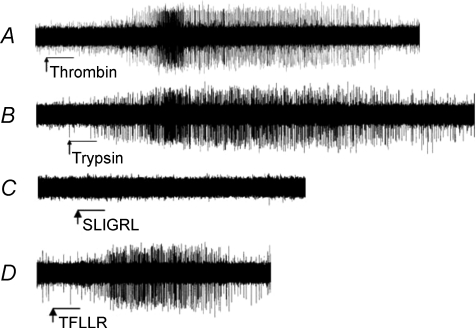
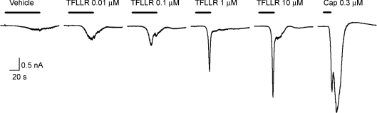
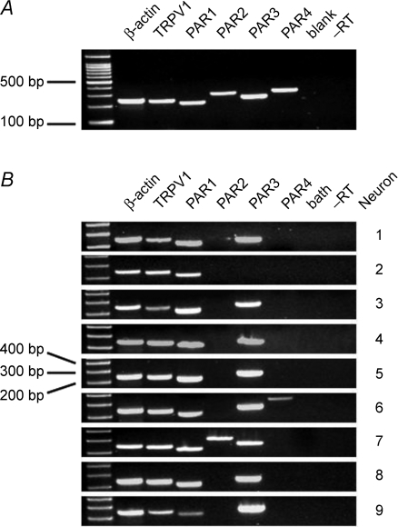
The nature of protease-activated receptors (PARs) capable of activating respiratory vagal C-fibres in the mouse was investigated. Infusing thrombin or trypsin via the trachea strongly activated vagal lung C-fibres with action potential discharge, recorded with the extracellular electrode positioned in the vagal sensory ganglion. The intensity of activation was similar to that observed with the TRPV1 agonist, capsaicin. This was mimicked by the PAR1-activating peptide TFLLR-NH(2), whereas the PAR2-activating peptide SLIGRL-NH(2) was without effect. Patch clamp recording on cell bodies of capsaicin-sensitive neurons retrogradely labelled from the lungs revealed that TFLLR-NH(2) consistently evokes a large inward current. RT-PCR revealed all four PARs were expressed in the vagal ganglia. However, when RT-PCR was carried out on individual neurons retrogradely labelled from the lungs it was noted that TRPV1-positive neurons (presumed C-fibre neurons) expressed PAR1 and PAR3, whereas PAR2 and PAR4 were rarely expressed. The C-fibres in mouse lungs isolated from PAR1(-/-) animals responded normally to capsaicin, but failed to respond to trypsin, thrombin, or TFLLR-NH(2). These data show that the PAR most relevant for evoking action potential discharge in vagal C-fibres in mouse lungs is PAR1, and that this is a direct neuronal effect.
Figures
References
-
- Amadesi S, Nie J, Vergnolle N, Cottrell GS, Grady EF, Trevisani M, Manni C, Geppetti P, McRoberts JA, Ennes H, Davis JB, Mayer EA, Bunnett NW. Protease-activated receptor 2 sensitizes the capsaicin receptor transient receptor potential vanilloid receptor 1 to induce hyperalgesia. J Neurosci. 2004;24:4300–4312. - PMC - PubMed
-
- Ashitani J, Mukae H, Arimura Y, Matsukura S. Elevated plasma procoagulant and fibrinolytic markers in patients with chronic obstructive pulmonary disease. Intern Med. 2002;41:181–185. - PubMed
-
- Barry GD, Le GT, Fairlie DP. Agonists and antagonists of protease activated receptors (PARs) Curr Med Chem. 2006;13:243–265. - PubMed
-
- Carr MJ, Schechter NM, Undem BJ. Trypsin-induced, neurokinin-mediated contraction of guinea pig bronchus. Am J Respir Crit Care Med. 2000;162:1662–1667. - PubMed
Publication types
MeSH terms
Substances
LinkOut - more resources
Full Text Sources
Other Literature Sources
Molecular Biology Databases
Miscellaneous