. 2010 Jun;19(7):815-29.
doi: 10.1177/0961203309360546. Epub 2010 Feb 8.

Spontaneous lupus-like syndrome in HLA-DQ2 transgenic mice with a mixed genetic background

Affiliations

Spontaneous lupus-like syndrome in HLA-DQ2 transgenic mice with a mixed genetic background

S Rashtak et al. Lupus. 2010 Jun.

Abstract

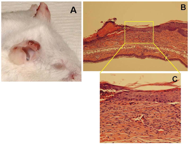
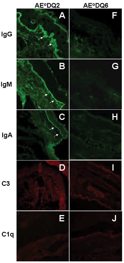
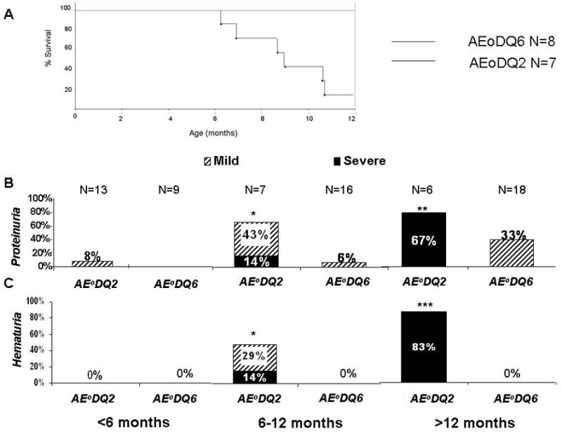
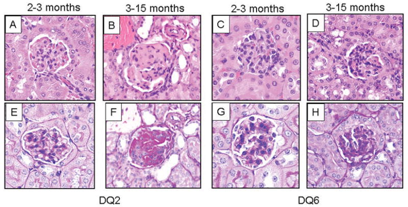
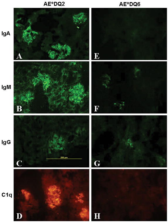
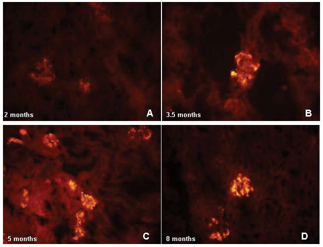
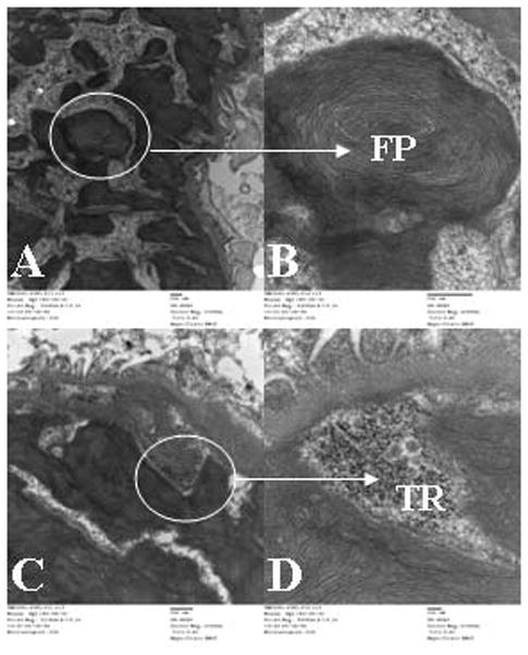
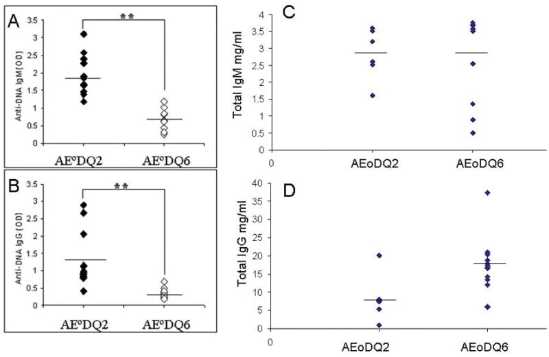
To investigate the role of HLA-DQ2 in the pathogenesis of associated immune disorders, we generated transgenic mice that expressed HLA-DQ2 in the absence of endogenous murine class II molecules (AE(0)DQ2). These AE(0)DQ2 mice with a mixed genetic background spontaneously developed skin lesions on their ears, whereas control AE(0)DQ6 genotype control mice (also with a mixed genetic background) did not. The skin lesions were characterized by deep subepidermal blistering with hydropic degeneration and lymphoid infiltration in the subepidermal area as determined by histopathology. Immunofluorescence analysis revealed thick band-like granular deposition of IgG, IgM, and a thin band of IgA deposition along the basement membrane. AE(0)DQ2 mice also developed significant and progressive hematuria and proteinuria as compared with the AE(0)DQ6 mice (p < 0.05). Histopathology showed immune complex deposits in the glomeruli of AE(0)DQ2 mice. Immunofluorescence analysis showed progressive mesangial and capillary wall deposition of IgA, IgM, IgG and C1q in the kidney. With electron microscopy, the deposits showed a 'fingerprint' substructure; and tubuloreticular structures were identified within endothelial cells. Conversely, these changes were not observed in AE(0)DQ6 mice. Serum anti-double stranded (ds)DNA IgM and IgG levels were also significantly elevated among AE(0)DQ2 mice compared with AE(0)DQ6 mice (p < 0.001). In conclusion, AE(0)DQ2 mice spontaneously develop an autoimmune lupus-like syndrome and are useful model for this disease. It remains to be determined whether genetic admixture played a role in the development of this systemic lupus erythematosus-like syndrome in HLA-DQ2 transgenic mice. Lupus (2010) 19, 815-829.


Figures

Figure 1
Figure 1
Chromosomal insertion sites of the DQ2 and DQ6 transgenes. (A) The Fluorescent in situ Hybridization (FISH) of metaphase chromosome slides from the spleens of AE°DQ2 mice (A) showed integration of probe on one chromosome (arrow). Samples from AE°DQ6 mice (B) had integration on three chromosomes (arrows). (C) Spectral karyotyping (SKY) revealed that the human HLA probe labeled in Vysis Spectrum Orange was integrated on one copy of mouse chromosome 8 in AE°DQ2 mice (arrow). (D) Evaluation of AE°DQ6 mice showed that the human HLA probe was integrated on both copies of mouse chromosome 2 and one copy of mouse chromosome 8 (arrows).
Figure 2
Figure 2
Expression of DQ2 and DQ6 molecules in AE°DQ2 and AE°DQ6 mice. (A–H) Reflective expression of cell surface DQ2 as determined by FACS analysis by splenocytes from five evaluated AE°DQ2 (A–D) and five AE°DQ6 (E–H) mice. A pan HLA-DQ, DR, DP antibody (TU39-green line) was used to detect DQ2 and DQ6. The isotype control (Mouse IgG2a) is the purple-filled area. Gates were set for B220+ cells (A, C, E, G) or CD11b+ cells (B, D, F, H), Splenocytes were cultured in vitro with media alone for 24 hours (A–B, E–F) or activated with LPS for 24 hours (C–D, G–H).
Figure 3
Figure 3
Development of cutaneous lesions in AE°DQ2 mice. (A) Erosions, ulcerations and crusting are present on the ears and muzzle. (B) H&E staining (10X) shows, thickening of the epidermis, hyperkeratosis as well as subepidermal blistering with hydropic degeneration and lymphoid infiltration in the subepidermal/dermal area (Reflective of 10 cutaneously affected mice). (C) Higher magnification (40x) shows the presence of vacuoles.
Figure 4
Figure 4
Direct immunofluorescence analysis of the skin. A thick band of IgG (A) and IgM (B) and a thin band of IgA (C) deposition with granular pattern was detected in the basement membrane of lesional ear in AE°DQ2 mice. There was no C3 (E) or C1q (D) deposition in the skin of AE°DQ2 mice (Reflective of five mice evaluated after 6 months of age). The AE°DQ6 mice did not show any IgG, IgM, IgA, C3 or C1q deposition in the skin (F–J).
Figure 5
Figure 5
Survival Analysis with Comparison of the percentage of positive urine blood and protein across genotype groups by age. Urine from AE°DQ2 mice and AE°DQ6 mice were subjected to strip analysis. Mice were categorized into three age groups: group one (0–6 months, mean age for AE°DQ2 mice versus AE°DQ6 mice was 4 versus 3 months in this group), group two (6–12 months, mean age for AE°DQ2 mice versus AE°DQ6 mice was 10 versus 11 months) and group three (>12 months, mean age for both AE°DQ2 and AE°DQ6 mice was15 months). (A) Kaplan Meier Curve for the survival rate of AEoDQ2 and AEo DQ6 mice. (B) Percentage of severe proteinuria (300 mg/dl or greater) and mild proteinuria (100mg/dl or greater, but less than 300 mg/dl). (C) Percentage of severe hematuria (3+ or greater) and mild hematuria (1+ or greater, but less than 3+). *P=0.02 for comparison of total hematuria and proteinuria, **P=0.001 for comparison of severe proteinuria, ***P<0.0001 for comparison of total hematuria and severe hematuria; Fisher’s exact test.
Figure 6
Figure 6
Histopathologic alterations in AE°DQ2 and AE°DQ6 mice. Reflective H&E stained kidney sections of AE°DQ2 mice between 2–3 months (A) of age seem completely normal. H&E stained section of the kidney of an AE°DQ2 mouse between 3–15 months of age (B). Glomeruli are hypocellular. Capillary loop lumens are markedly compromised due to subendothelial deposition of immune complex deposits. Tubules and interstitium are unremarkable. H&E stained sections of AE°DQ6 kidneys from 2–3 months of age (C) and 3–15 months of age (D), revealed no significant histopathologic alterations of glomeruli, tubules, interstitial, or vessels. PAS stained kidney section of a 2 month old AE°DQ2 mouse showing minimal mesangial hypercellularity (E). The tubules, interstitium, and vessels are unremarkable. (F) PAS stained section of AE°DQ2 mouse at 14 months of age: Glomeruli show massive subendothelial immune complex deposits. Patchy interstitial lymphocytic infiltrates are identified. Original magnification, ×40. PAS stained sections of AEoDQ6 kidneys from mice aged 2 months (G) and 14 months (H) appear normal.
Figure 7
Figure 7
Direct immunofluorescent analysis of kidney specimens for presence of IgA, IgM, IgG and C1q in AE°DQ2 (at 11 months of age) and AE°DQ6 (at 12 months of age) mice. (A–D) Glomeruli of AE°DQ2 mice demonstrated strong diffuse granular mesangial and capillary wall staining for IgA (A), IgM (B), weak granular mesangial staining for IgG (C), and strong diffuse granular and capillary wall staining for C1q (D). (E–H) By contrast, AE°DQ6 mice showed no significant staining for IgA (E), IgG (G), C1q (H), and only trace granular mesangial staining for IgM (F). Original magnification, ×40.
Figure 8
Figure 8
Progression of complement deposition by age in AE°DQ2 mice. (A) At 2 months of age C1q was present in low intensity in the kidney, however the intensity increased over time as the mice aged (B) 3.5 months of age; (C) 5 months of age; (D) 8 months of age.
Figure 9
Figure 9
Electron microscopic studies. Electron micrographs were taken of kidneys from an AE°DQ2 mouse of 11 months of age. (A) Massive mesangial and subendothelial immune complex-type deposits were present. The large subendothelial deposits markedly compromised peripheral capillary loop lumens, and were reminiscent of the “wire loop” lesions observed in lupus glomerulonephritis (Direct magnification, ×11,000). (B) On high power magnification the immune complex deposits had a vaguely paracrystalline appearance with a “fingerprint” (FP) substructure (Direct magnification, ×42,000). (C) Tubuloreticular (TR) structures were identified within endothelial cells (Direct magnification, ×21,000). (D) TR on high power (Direct magnification, ×67,000).
Figure 10
Figure 10
Comparison of anti-DNA antibody levels in the sera of AE°DQ2 and AE°DQ6 mice. (A) Anti-DNA IgM (OD) and (B) Anti-DNA IgG (OD) were measured by ELISA method in the serum of AE°DQ2 (Mean age 10.3, range 8–14 months, N=12) and AE°DQ6 (Mean age 11.6, range 11–12 months, N=9) mice. Horizontal bars represent the means. **P<0.001; Wilcoxon rank sum test. (C–D) Total IgM and total IgG levels in sera of AEoDQ2 and AEoDQ6 mice.

Similar articles

Cited by

References

    1. Martin-Villa JM, Martinez-Laso J, Moreno-Pelayo MA, Castro-Panete MJ, Martinez-Quiles N, Alvarez M, de Juan MD, Gomez-Reino JJ, Arnaiz-Villena A. Differential contribution of HLA-DR, DQ, and TAP2 alleles to systemic lupus erythematosus susceptibility in Spanish patients: role of TAP2*01 alleles in Ro autoantibody production. Ann Rheum Dis. 1998;57:214–9. - PMC - PubMed
    1. Vargas-Alarcon G, Salgado N, Granados J, Gomez-Casado E, Martinez-Laso J, Alcocer-Varela J, Arnaiz-Villena A, Alarcon-Segovia D. Class II allele and haplotype frequencies in Mexican systemic lupus erythematosus patients: the relevance of considering homologous chromosomes in determining susceptibility. Hum Immunol. 2001;62:814–20. - PubMed
    1. Tjernstrom F, Hellmer G, Nived O, Truedsson L, Sturfelt G. Synergetic effect between interleukin-1 receptor antagonist allele (IL1RN*2) and MHC class II (DR17,DQ2) in determining susceptibility to systemic lupus erythematosus. Lupus. 1999;8:103–8. - PubMed
    1. Lambert AP, Gillespie KM, Thomson G, Cordell HJ, Todd JA, Gale EA, Bingley PJ. Absolute risk of childhood-onset type 1 diabetes defined by human leukocyte antigen class II genotype: a population-based study in the United Kingdom. J Clin Endocrinol Metab. 2004;89:4037–43. - PubMed
    1. Segni M, Pani MA, Pasquino AM, Badenhoop K. Familial clustering of juvenile thyroid autoimmunity: higher risk is conferred by human leukocyte antigen DR3-DQ2 and thyroid peroxidase antibody status in fathers. J Clin Endocrinol Metab. 2002;87:3779–82. - PubMed

Publication types