. 2010 Mar-Apr;3(3-4):224-35.
doi: 10.1242/dmm.004226. Epub 2010 Feb 8.

Complementary interplay between matrix metalloproteinase-9, vascular endothelial growth factor and osteoclast function drives endochondral bone formation

Affiliations

Complementary interplay between matrix metalloproteinase-9, vascular endothelial growth factor and osteoclast function drives endochondral bone formation

Nathalie Ortega et al. Dis Model Mech. 2010 Mar-Apr.

Abstract

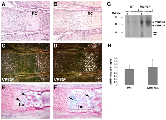
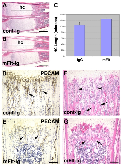
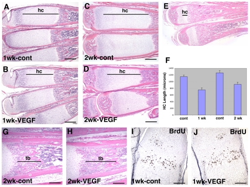
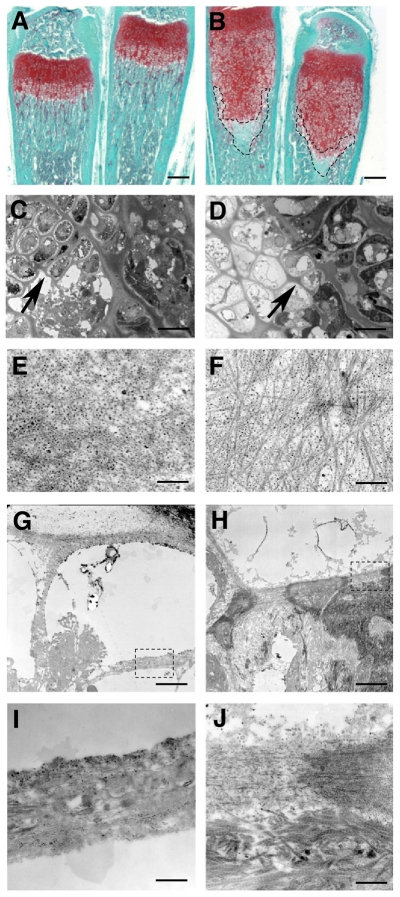
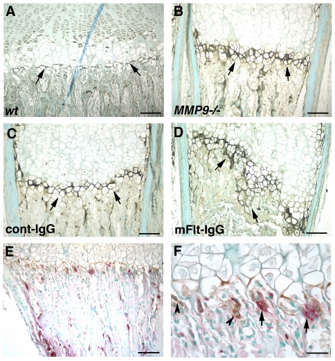
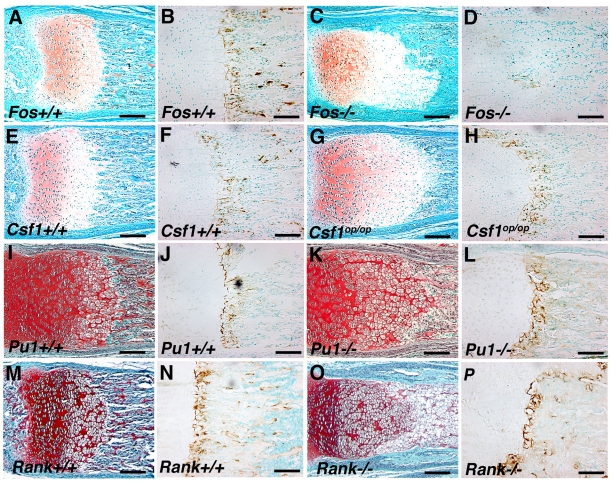
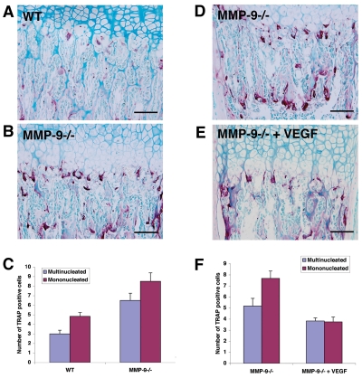
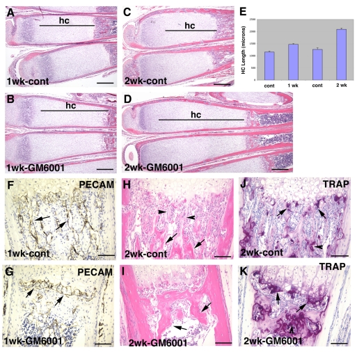
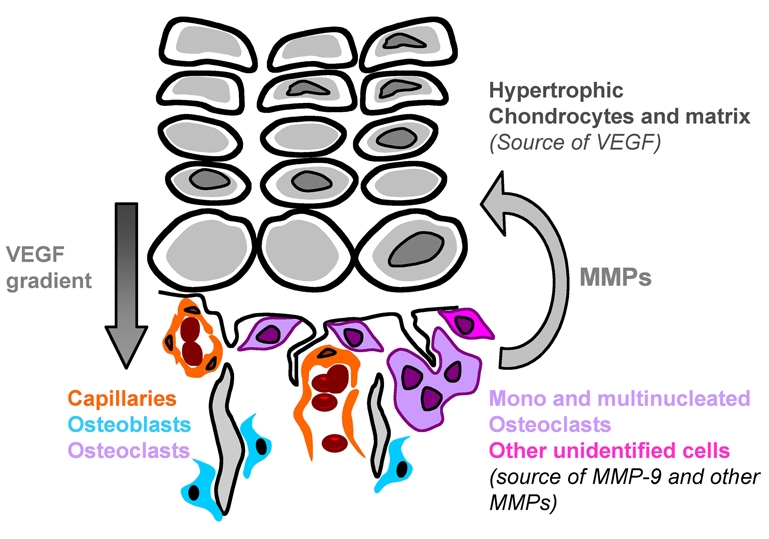
Long bone development depends on endochondral bone formation, a complex process requiring exquisite balance between hypertrophic cartilage (HC) formation and its ossification. Dysregulation of this process may result in skeletal dysplasias and heterotopic ossification. Endochondral ossification requires the precise orchestration of HC vascularization, extracellular matrix remodeling, and the recruitment of osteoclasts and osteoblasts. Matrix metalloproteinase-9 (MMP-9), vascular endothelial growth factor (VEGF) and osteoclasts have all been shown to regulate endochondral ossification, but how their function interrelates is not known. We have investigated the functional relationship among these regulators of endochondral ossification, demonstrating that they have complementary but non-overlapping functions. MMP-9, VEGF and osteoclast deficiency all cause impaired growth plate ossification resulting in the accumulation of HC. VEGF mRNA and protein expression are increased at the MMP-9-/- growth plate, and VEGF activity contributes to endochondral ossification since sequestration of VEGF by soluble receptors results in further inhibition of growth plate vascularization and ossification. However, VEGF bioavailability is still limited in MMP-9 deficiency, as exogenous VEGF is able to rescue the MMP-9-/- phenotype, demonstrating that MMP-9 may partially, but not fully, regulate VEGF bioavailability. The organization of the HC extracellular matrix at the MMP-9-/- growth plate is altered, supporting a role for MMP-9 in HC remodeling. Inhibition of VEGF impairs osteoclast recruitment, whereas MMP-9 deficiency leads to an accumulation of osteoclasts at the chondro-osseous junction. Growth plate ossification in osteoclast-deficient mice is impaired in the presence of normal MMP-9 expression, indicating that other osteoclastic functions are also necessary. Our data delineate the complementary interplay between MMP-9, VEGF and osteoclast function that is necessary for normal endochondral bone formation and provide a molecular framework for investigating the molecular defects contributing to disorders of endochondral bone formation.


Figures

Fig. 1.
Fig. 1.
VEGF is expressed in the MMP-9−/− mice. (A–D) Bright-field (A,B) and dark-field (C,D) photographs of WT (A,C) and MMP-9−/− (B,D) sections of 1-week-old metatarsal growth plates hybridized with a Vegf antisense probe. Vegf is expressed in a subpopulation of hypertrophic chondrocytes (hc). (E,F) Immunostaining of tissue sections of 1-week-old WT (E) and MMP-9−/− (F) growth plates with VEGF antibody. VEGF protein is found in the ECM surrounding the hypertrophic chondrocytes (arrows). (G) Western blot of protein extracts from 2-week-old WT and MMP-9−/− growth plate HC probed with an anti-VEGF antibody showing that the major VEGF isoform expressed is VEGF164. Note the smaller forms of VEGF migrating around 28 kDa in both WT and MMP-9−/− HC with intact perichondrium (arrows). (H) Quantification by ELISA of VEGF protein secreted into the medium by cultured 2-week-old WT and MMP-9−/− growth plate ossification fronts. Bars, 200 μm (A–F).
Fig. 2.
Fig. 2.
Inhibition of VEGF activity causes further accumulation of HC in the MMP-9−/− growth plate. (A,B) Hematoxylin and eosin (H&E)-stained sections of 1-week-old MMP-9−/− mice treated for 10 days with either control IgG (A) or mFlt(1–3)-IgG (B) showing lengthening of the HC zone (hc) in the treated mice. (C) Bar graphs of the mean and standard deviation of the HC zone lengths of six control and mFlt-treated bones; the differences between control and treated samples were significant (P<0.05). (D,E) Immunostaining of tissue sections with PECAM-1 antibody showing metaphyseal vessels running parallel to hypertrophic chondrocyte columns in the control-IgG-treated mice (D, arrows) and a reduced number of these vessels in the mFlt(1–3)-IgG-treated mice (E, arrows). (F,G) H&E-stained sections showing primary (F, arrowheads) and secondary (F, arrows) trabeculae in the control-IgG-treated mice, and thickened and shortened secondary trabeculae (G, arrows) in the mFlt(1–3)-IgG-treated mice. Bars, 400 μm (A,B); 100 μm (D–G).
Fig. 3.
Fig. 3.
Treatment with VEGF rescues endochondral ossification defects in MMP-9 −/− mice. (A–D) H&E-stained sections of metatarsals of 1-week-old MMP-9−/− mice treated for 1 week (A,B) or 2 weeks (C,D) with either vehicle or recombinant human VEGF165, showing shortening of the HC zone (hc) in VEGF-treated mice (B,D) compared with vehicle-treated mice (A,C). (E) H&E-stained section of 3-week-old WT metatarsals showing the size of the normal HC zone (hc). (F) Bar graphs of the mean and standard deviation of the HC zone lengths of six vehicle- and VEGF-treated bones after 1 and 2 weeks of treatment; the differences between control and treated samples at each time point were significant (P<0.01). (G,H) H&E-stained sections of the metatarsals of MMP-9−/− mice treated for 2 weeks with either vehicle (G) or VEGF (H) showing lengthening of the trabecular bone region (tb) in VEGF-treated mice. (I,J) BrdU labeling of proliferating cells in the metatarsal growth plates of 1-week-old MMP-9−/− mice treated for 1 week with VEGF. BrdU-positive cells stain brown. Bars, 400 μm (A–D); 200 μm (G,H); 100 μm (I,J).
Fig. 4.
Fig. 4.
Altered hypertrophic cartilage ECM in MMP-9 null growth plates. (A,B) Safranin-O staining of 3-week-old metatarsals from WT (A) and MMP-9−/− (B) mice, showing a decrease in the staining of the lower portion of the hypertrophic chondrocyte area in MMP-9−/− mice (outlined by dotted lines). (C,D) Semi-thin sections of Epon-embedded ossification fronts from WT (C) and MMP-9−/− (D) mice, stained with Toluidine Blue, showing the presence of hypertrophic chondrocytes in the last rows at the ossification fronts in MMP-9−/− mice compared with WT mice. The longitudinal septae of the lacunae surrounding the last rows of hypertrophic chondrocytes are thicker in the MMP-9−/− mice (D, arrow) than in the WT mice (C, arrow). (E,F) Transmission electron micrographs of the areas indicated by arrows in C and D, showing the ultrastructure of the ECM in these areas. The ECM in these septae in WT mice is rich in proteoglycans, as indicated by numerous black dots, and collagen fibers are sparse (E). By contrast, in MMP-9−/− mice, the ECM of these septae contains dense collagen fibers and fewer black dots, indicating a decrease in proteoglycan content, which was also observed with Safranin-O staining in A and B. (G,H) Transmission electron micrographs showing the ultrastructure of the ECM of the last transverse septa in WT (G) and MMP-9−/− (H) mice. The boxed areas are shown under higher magnification in I and J. Transverse septae in the MMP-9−/− samples (J) show more collagen fibers and there is an accumulation of collagen type I fibers under the septae compared with the transverse septae in WT samples (I). Bars, 500 μm (A,B); 50 μm (C,D); 20 nm (E,F); 16 μm (G,H); 2 μm (I,J).
Fig. 5.
Fig. 5.
MMP-9 deficiency and inhibition of VEGF alter the accumulation of cleaved collagen at the growth plate. (A–D) Immunostaining with an antibody against cleaved fragments of type II collagen on sections of metatarsals of 2-week-old WT (A) and MMP-9−/− (B) mice, and of sections of 1-week-old MMP-9−/− mice treated with cont-IgG (C) or mFlt(1–3)-IgG (D) for 1 week, showing the presence of cleaved collagen at a thin line along the cartilage-bone junction in WT mice (A, arrows), a larger zone of cleaved collagen in MMP-9−/− mice (B, arrows), and a larger zone with an irregular border in MMP-9−/− mice treated with mFlt(1–3)-IgG (D, arrows). (E,F) Immunostaining for MMP-9 and substrate staining for tartrate-resistant acidic phosphatase (TRAP) activity on the same tissue sections from WT growth plates showing MMP-9-positive cells that are both TRAP positive (F, arrows) and TRAP negative (F, arrowheads). Bars, 100 μm (A–E); 32 μm (F).
Fig. 6.
Fig. 6.
Osteopetrotic mice have growth plate abnormalities with normal expression of MMP-9. Histological sections of metatarsal growth plates from c-Fos+/+ (A), c-Fos−/− (C), Csf1+/+ (E), Csf1op/op (G), PU.1+/+ (I), PU.1−/− (K), Rank+/+ (M) and Rank−/− (O) mice showing enlarged HC zones in the mutant growth plates. Immunostaining for MMP-9 showed similar expression of MMP-9 in the Csf1+/+ (F), Csf1op/op (H), PU.1+/+ (J), PU.1−/− (L), Rank+/+ (N) and Rank −/− (P) growth plates. The c-Fos−/− growth plate showed decreased immunostaining for MMP-9 (D) compared with the control c-Fos+/+ growth plate (B). Bars, 100 μm.
Fig. 7.
Fig. 7.
Osteoclast recruitment to the chondro-osseous junction. (A,B) TRAP staining of tissue sections showing more TRAP+ cells at the chondro-osseous junction in MMP-9−/− growth plates (B) compared with WT growth plates (A). (C) Quantification of mononuclear and multinuclear TRAP+ cells at the WT and MMP-9−/− growth plates. (D,E) TRAP staining of tissue sections showing a reduced number of TRAP+ cells at the chondro-osseous junction in MMP-9−/− growth plates treated with VEGF (E) compared with untreated MMP-9−/− growth plates (D). (F) Quantification of mononuclear and multinuclear TRAP+ cells at the untreated and VEGF-treated MMP-9−/− growth plates. For C and F, mononucleated and multinucleated TRAP+ cells were counted under the microscope at a 40× magnification and expressed as the total number of TRAP+ cells along the total length of the chondro-osseous junction. The counting was performed on a minimum of three mice per group, using both hind limbs, and on three to six different sections, with three metatarsals per section, for each hind limb. The results are expressed as mean ± standard error of the mean (S.E.M.). The results were statistically significant (P<0.01) between WT and MMP-9−/− samples for both cell populations in C, and between VEGF treated and untreated samples for the mononuclear population in F. The results were not statistically significant between VEGF treated and untreated samples for the multinuclear population in F.
Fig. 8.
Fig. 8.
MMP expression at the growth plate. Dark-field photographs of tissue sections of metatarsals from 1-week-old WT (A,C) and MMP-9−/− mice (B,D) hybridized with antisense probes against MMP-13 (A,B) and MMP-14 (C,D). MMP-13 is expressed in cells at the cartilage-bone junction, consistent with the previously reported expression in terminal hypertrophic chondrocytes (A,B, arrowheads) and in the metaphysis (A,B, asterisks), in both WT and MMP-9−/− mice. In addition, MMP-13 is also expressed in some lower hypertrophic chondrocytes in MMP-9−/− mice (B, arrows). MMP-14 is expressed in cells at the cartilage-bone junction (C,D, arrowheads) and the metaphysis (C,D, asterisks). It is also expressed in the perichondrium (C,D, arrows). Bars, 200 μm.
Fig. 9.
Fig. 9.
Inhibition of MMP activity inhibits residual endochondral ossification at the MMP-9−/− growth plate. (A–D) H&E-stained sections of metatarsals of 1-week-old MMP-9−/− mice treated for 1 week (A,B) or 2 weeks (C,D) with either vehicle or the MMP inhibitor GM6001, showing further lengthening of the HC (hc) zone in the GM6001-treated mice (B,D) compared with the vehicle-treated mice (A,C). (E) Bar graphs of the mean and standard deviation of the HC zone lengths of six control and GM6001-treated bones after 1 and 2 weeks of treatment; the differences between control and treated samples at each time point were statistically significant (P<0.01). (F,G) Immunostaining of tissue sections with PECAM-1 antibody showing metaphyseal vessels running parallel to hypertrophic chondrocyte columns in the vehicle-treated mice (F, arrows) and dilated vessels running perpendicular to the hypertrophic chondrocyte columns in the GM6001-treated mice (G, arrows). (H,I) H&E-stained sections showing primary (H, arrowheads) and secondary (H, arrows) trabeculae in the vehicle-treated mice, and a mass of bone instead of secondary trabeculae in the GM6001-treated mice (I, arrows). (J,K) TRAP staining of tissue sections showing the presence of TRAP+ cells at the cartilage-bone junction (arrows) and on trabecular bone surfaces (arrowheads) in both vehicle-treated (J) and GM6001-treated (K) mice. Bars, 400 μm (A–D); 100 μm (F–K).
Fig. 10.
Fig. 10.
Complementary functions of VEGF, MMP-9 and osteoclasts at the growth plate. At the HC-bone junction, MMP-9 is secreted by mononucleated and multinucleated osteoclasts, as well as by other non-osteoclastic cells. MMP-9 can degrade the ECM leading to the release of VEGF sequestered in the HC. However, MMP-9 also has functions besides regulating VEGF bioavailability, such as ECM degradation and remodeling. VEGF can act on endothelial cells, as well as on mononucleated osteoclasts and osteoblasts. Besides MMP-9, osteoclasts also secrete other MMPs that can also cleave the ECM.

Similar articles

Cited by

References

    1. Aldridge SE, Lennard TW, Williams JR, Birch MA. (2005). Vascular endothelial growth factor receptors in osteoclast differentiation and function. Biochem Biophys Res Commun. 335, 793–798 - PubMed
    1. Arikawa-Hirasawa E, Watanabe H, Takami H, Hassell JR, Yamada Y. (1999). Perlecan is essential for cartilage and cephalic development. Nat Genet. 23, 354–358 - PubMed
    1. Bergers G, Brekken R, McMahon G, Vu TH, Itoh T, Tamaki K, Tanzawa K, Thorpe P, Itohara S, Werb Z, et al. (2000). Matrix metalloproteinase-9 triggers the angiogenic switch during carcinogenesis. Nat Cell Biol. 2, 737–744 - PMC - PubMed
    1. Billinghurst RC, Dahlberg L, Ionescu M, Reiner A, Bourne R, Rorabeck C, Mitchell P, Hambor J, Diekmann O, Tschesche H, et al. (1997). Enhanced cleavage of type II collagen by collagenases in osteoarthritic articular cartilage. J Clin Invest 99, 1534–1545 - PMC - PubMed
    1. Bischof P, Truong K, Campana A. (2003). Regulation of trophoblastic gelatinases by proto-oncogenes. Placenta 24, 155–163 - PubMed

Publication types

MeSH terms