. 2010 Mar-Apr;3(3-4):181-93.
doi: 10.1242/dmm.004440. Epub 2010 Feb 8.

Lkb1 inactivation is sufficient to drive endometrial cancers that are aggressive yet highly responsive to mTOR inhibitor monotherapy

Affiliations

Lkb1 inactivation is sufficient to drive endometrial cancers that are aggressive yet highly responsive to mTOR inhibitor monotherapy

Cristina M Contreras et al. Dis Model Mech. 2010 Mar-Apr.

Abstract

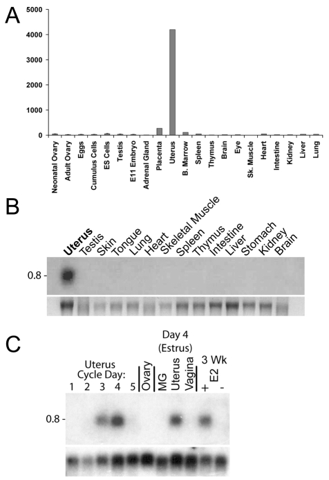
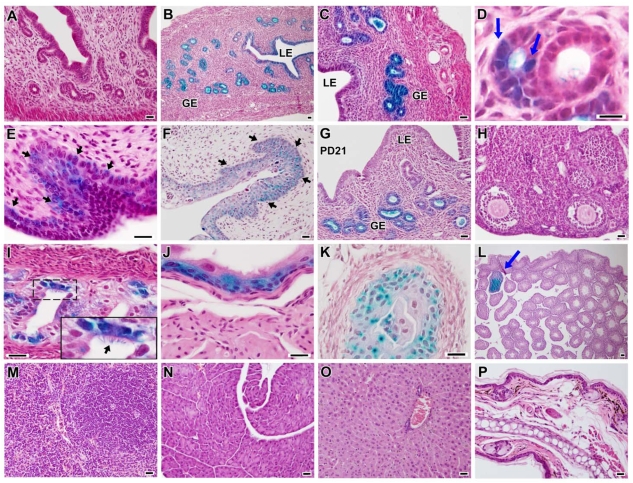
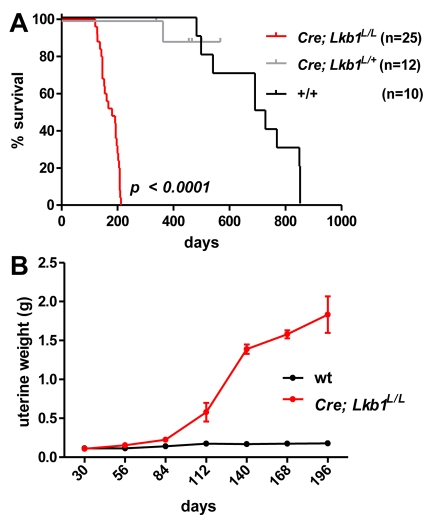
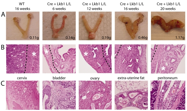
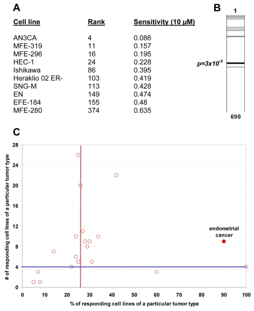
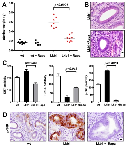
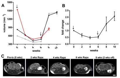
Endometrial cancer--the most common malignancy of the female reproductive tract--arises from the specialized epithelial cells that line the inner surface of the uterus. Although significant advances have been made in our understanding of this disease in recent years, one significant limitation has been the lack of a diverse genetic toolkit for the generation of mouse models. We identified a novel endometrial-specific gene, Sprr2f, and developed a Sprr2f-Cre transgene for conditional gene targeting within endometrial epithelium. We then used this tool to generate a completely penetrant Lkb1 (also known as Stk11)-based mouse model of invasive endometrial cancer. Strikingly, female mice with homozygous endometrial Lkb1 inactivation did not harbor discrete endometrial neoplasms, but instead underwent diffuse malignant transformation of their entire endometrium with rapid extrauterine spread and death, suggesting that Lkb1 inactivation was sufficient to promote the development of invasive endometrial cancer. Mice with heterozygous endometrial Lkb1 inactivation only rarely developed tumors, which were focal and arose with much longer latency, arguing against the idea--suggested by some prior studies--that Lkb1 is a haploinsufficient tumor suppressor. Lastly, the finding that endometrial cancer cell lines were especially sensitive to the mTOR (mammalian target of rapamycin) inhibitor rapamycin prompted us to test its efficacy against Lkb1-driven endometrial cancers. Rapamycin monotherapy not only greatly slowed disease progression, but also led to striking regression of pre-existing tumors. These studies demonstrate that Lkb1 is a uniquely potent endometrial tumor suppressor, but also suggest that the clinical responses of some types of invasive cancers to mTOR inhibitors may be linked to Lkb1 status.


Figures

Fig. 1.
Fig. 1.
Identification of Sprr2f as an estrogen-dependent gene that is specifically expressed in the uterus. (A) Digital northern representation, per Affymetrix 430 2.0 array data, of Sprr2f expression in total RNA from various tissues and developmental stages. The Y-axis corresponds to the relative expression levels of the Sprr2f probe set (1449833_at) across the samples shown. Tissues were obtained from adult mice unless specified otherwise. (B) Northern analysis of total RNA using an Sprr2f 3′-UTR probe demonstrates that Sprr2f (0.8 kb transcript) is expressed only in the uterus and in no other tissues obtained from adult female mice. A probe corresponding to the 3′-UTR was selected to avoid cross-hybridization with other Sprr2 members, which are highly conserved. Bottom panel=loading control. (C) Northern analysis of total RNA using 3′-UTR probe, showing that Sprr2f expression is cyclical in relation to estrus and is induced by estrogen; MG=mammary gland. Uterus cycle days D1–D5 correspond to diestrus, proestrus, early estrus, estrus and postestrus. The last two lanes represent uteri from 3-week-old mice injected with estrogen or vehicle. Bottom panel=loading control.
Fig. 2.
Fig. 2.
Cre activity in gonads and other tissues in Sprr2f-Cre; R26R reporter mice. Tissues were fixed in formalin, stained with X-gal, and sectioned. All tissues are from adult mice (6-week old), except where noted. (A) Uterus; the uterus of a R26R non-transgenic sibling (negative control) shows an absence of Cre activity. (B,C) Uterus (higher magnification); the uterus shows efficient and specific Cre-mediated recombination within endometrial epithelium. LE=luminal epithelium, GE=glandular epithelium. (D) Uterus; a close-up of shows a mosaic pattern of recombination where only some cells have undergone Cre-mediated recombination (arrows). (E) Cervix; the squamocolumnnar junction (arrows show efficient recombination in the superficial and reserve layer). (F) Vaginal fornix. (G) Uterus from a 3-week-old mouse (postnatal day 21). Recombination was already efficient in the glandular epithelium in these prepubertal animals (it ranged from ~30 to 50%); the field shown represents a region of efficient recombination. (H) Ovary. (I) Oviduct; the inset shows specific recombination in ciliated epithelial cells (arrows). (J) Bladder. (K) Ureter. (L) Testis; the arrow shows a rare tubule with focal Cre activity. (M) Spleen. (N) Pancreas. (O) Liver. (P) Ear with skin, cartilage and connective tissue. Bars, 20 μm (A–C,E–J,L–P); 40 μm (D,K).
Fig. 3.
Fig. 3.
Homozygous but not heterozygous inactivation of Lkb1 results in accelerated mortality owing to endometrial cancer. (A) Survival curve; P<0.0001 for both Sprr2f-Cre; Lkb1L/L versus wild-type controls and Sprr2f-Cre; Lkb1L/L versus Sprr2f-Cre; Lkb1L/+ sibling heterozygotes. The +/+ curve represents non-sibling wild-type animals of the same genetic background. Statistical significance was calculated with the log-rank test. (B) Uterine weights of Sprr2f-Cre; Lkb1L/L and wild-type animals, n=1–5 uteri per time point. Error bars=standard error of the mean (S.E.M.).
Fig. 4.
Fig. 4.
Stereotypical endometrial cancer initiation and progression in Sprr2f-Cre; Lkb1L/L mice. (A) Gross pictures of intact uteri at 6 to 20 weeks of age; a control (wild-type) uterus at 16 weeks is shown on the left. The weight for each uterus is shown in the lower right corner. (B) Microscopic analysis shows a lack of invasion at 6 weeks and progressive infiltration after 12 weeks. The endometrial-myometrial interface is shown by a dashed line, with the myometrial layer marked by an asterisk. Note: distinctive features of Lkb1-deficient endometrial tumors include their extremely well-differentiated histology and the absence of a morphologically distinctive in situ precursor. Infiltration of glands into the myometrium (which does not normally harbor endometrial glands) serves as definitive histological evidence of invasion (Contreras et al., 2008a). (C) Growth of invasive endometrial adenocarcinoma in characteristic anatomic locations. At each site, the tumor retains a well-differentiated appearance, consisting of well-defined interspersed glands associated with abundant stroma.
Fig. 5.
Fig. 5.
Endometrial cancer cell lines are especially sensitive to rapamycin. (A) Rank order of each endometrial cancer cell line (n=10) among a total of 690 cancer cell lines in the study. Sensitivity was calculated as the fraction of viable cells relative to untreated controls following treatment with 10 μM rapamycin. The P value was calculated as described in the methods section. (B) Graphical representation of ranks. Each horizontal bar represents the rank order position of a single endometrial cancer cell line among the 690 cell lines, except for the thick bar, which represents the median rank. Note that nine of the ten cell lines are in the top half. (C) Tumor type rapamycin sensitivity chart. Each circle corresponds to a tumor type (e.g. pancreatic, lung, breast). The vertical red line indicates the overall sensitivity of all cell lines (n=690) to the compound; i.e. 26% of human tumor-derived cell lines were sensitive to rapamycin (the criteria for sensitivity was >50% killing at a high concentration of the drug). The horizontal blue line at 4 on the y-axis is an empirically-derived gate indicating that, for any sensitivity to be considered meaningful, it has to occur in >4 cell lines of a particular tumor type. Tumor types in the top right-hand quadrant are thus more sensitive to the drug. The endometrial adenocarcinoma group at 90% (highlighted in red) includes all of the endometrial adenocarcinoma cell lines (n=10).
Fig. 6.
Fig. 6.
Rapamycin monotherapy slows the progression of Lkb1-deficient endometrial adenocarcinomas. (A) Uterine weights following 4 weeks of rapamycin monotherapy. Samples are wild-type mice injected with vehicle only (wt; n=9), wild-type mice treated with rapamycin (wt+Rapa; n=5), Sprr2f-Cre; Lkb1L/L mice injected with vehicle only (Lkb1; n=7), and Sprr2f-Cre; Lkb1L/L mice treated with rapamycin (Lkb1+Rapa; n=9). (B) The tumor burden is reduced greatly by rapamycin therapy but, histologically, the few residual tumor glands closely resemble the tumor glands in untreated animals. (C) Ki67, TUNEL and p-S6K positivity in endometrial epithelium. Bars represent relative counts in tissue sections. (D) Representative regions of tissue sections that were immunohistochemically stained for p-S6K and counterstained with hematoxylin. Bars, 100 μm (B,D).
Fig. 7.
Fig. 7.
Rapamycin monotherapy leads to dramatic responses even in animals with very advanced tumors. (A) Uterine volumes (in cubic millimeters), as determined by serial MRI performed every two weeks. Each line represents a single animal. Death, or severe morbidity requiring euthanasia prior to the next scheduled MRI scan, is indicated by a dagger (†) symbol. (B) Relative fold-change of uterine volume, expressed as an average for all three animals (the same animals as shown in panel A). Error bars=S.E.M. For panels A and B, the first arrow marks the initiation of therapy, and the second arrow marks cessation of therapy. (C) Representative MRI images of axial sections showing the effect of rapamycin therapy. Top=ventral. In each image, a dashed white line indicates the uterine margin. The animal shown corresponds to the animal represented by a black line in panel A. Bar, 5 mm.

Similar articles

Cited by

References

    1. Akbay EA, Contreras CM, Perera SA, Sullivan JP, Broaddus RR, Schorge JO, Ashfaq R, Saboorian H, Wong KK, Castrillon DH. (2008). Differential roles of telomere attrition in type I and II endometrial carcinogenesis. Am J Pathol. 173, 536–544 - PMC - PubMed
    1. Alessi DR, Sakamoto K, Bayascas JR. (2006). Lkb1-dependent signaling pathways. Annu Rev Biochem. 75, 137–163 - PubMed
    1. Bajic VB, Tan SL, Chong A, Tang S, Strom A, Gustafsson JA, Lin CY, Liu ET. (2003). Dragon ERE Finder version 2, A tool for accurate detection and analysis of estrogen response elements in vertebrate genomes. Nucleic Acids Res. 31, 3605–3607 - PMC - PubMed
    1. Barakat RR, Grigsby PW, Sabbatini P, Zaino RJ. (2000). Corpus: Epithelial Tumors (in Principles and Practice of Gynecologic Oncology). Philadelphia: Lippincott Williams and Wilkins
    1. Bissler JJ, McCormack FX, Young LR, Elwing JM, Chuck G, Leonard JM, Schmithorst VJ, Laor T, Brody AS, Bean J, et al. (2008). Sirolimus for angiomyolipoma in tuberous sclerosis complex or lymphangioleiomyomatosis. N Engl J Med. 358, 140–151 - PMC - PubMed

Publication types

MeSH terms