

Tapping into the glial reservoir: cells committed to remaining uncommitted
- PMID: 20142420
- PMCID: PMC2819683
- DOI: 10.1083/jcb.200905111
Tapping into the glial reservoir: cells committed to remaining uncommitted
Abstract
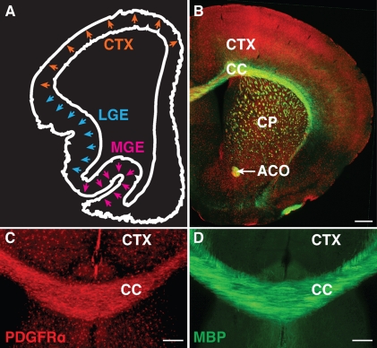
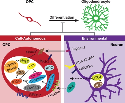
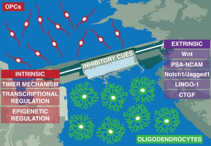
The development and maturation of the oligodendrocyte requires a series of highly orchestrated events that coordinate the proliferation and differentiation of the oligodendrocyte precursor cell (OPC) as well as the spatiotemporal regulation of myelination. In recent years, widespread interest has been devoted to the therapeutic potential of adult OPCs scattered throughout the central nervous system (CNS). In this review, we highlight molecular mechanisms controlling OPC differentiation during development and the implication of these mechanisms on adult OPCs for remyelination. Cell-autonomous regulators of differentiation and the heterogeneous microenvironment of the developing and the adult CNS may provide coordinated inhibitory cues that ultimately maintain a reservoir of uncommitted glia.
Figures
Similar articles
-
Schwann cell remyelination of the central nervous system: why does it happen and what are the benefits?Open Biol. 2021 Jan;11(1):200352. doi: 10.1098/rsob.200352. Epub 2021 Jan 27. Open Biol. 2021. PMID: 33497588 Free PMC article. Review.
-
Early proliferation does not prevent the loss of oligodendrocyte progenitor cells during the chronic phase of secondary degeneration in a CNS white matter tract.PLoS One. 2013 Jun 11;8(6):e65710. doi: 10.1371/journal.pone.0065710. Print 2013. PLoS One. 2013. PMID: 23776532 Free PMC article.
-
Evidence that perinatal and adult NG2-glia are not conventional oligodendrocyte progenitors and do not depend on axons for their survival.Mol Cell Neurosci. 2003 Aug;23(4):544-58. doi: 10.1016/s1044-7431(03)00176-3. Mol Cell Neurosci. 2003. PMID: 12932436
-
Engineering biomaterial microenvironments to promote myelination in the central nervous system.Brain Res Bull. 2019 Oct;152:159-174. doi: 10.1016/j.brainresbull.2019.07.013. Epub 2019 Jul 12. Brain Res Bull. 2019. PMID: 31306690 Review.
-
Cytology and lineage of NG2-positive glia.J Neurocytol. 2002 Jul-Aug;31(6-7):457-67. doi: 10.1023/a:1025735513560. J Neurocytol. 2002. PMID: 14501216 Review.
Cited by
-
Characterization of Glial Populations in the Aging and Remyelinating Mouse Corpus Callosum.Neurochem Res. 2022 Sep;47(9):2826-2838. doi: 10.1007/s11064-022-03676-z. Epub 2022 Jul 20. Neurochem Res. 2022. PMID: 35859078
-
Silencing or knocking out the Na(+)/Ca(2+) exchanger-3 (NCX3) impairs oligodendrocyte differentiation.Cell Death Differ. 2012 Apr;19(4):562-72. doi: 10.1038/cdd.2011.125. Epub 2011 Sep 30. Cell Death Differ. 2012. PMID: 21959935 Free PMC article.
-
Epigenetic regulation of human-specific gene expression in the prefrontal cortex.BMC Biol. 2023 May 24;21(1):123. doi: 10.1186/s12915-023-01612-3. BMC Biol. 2023. PMID: 37226244 Free PMC article.
-
Modulation of oligodendrocyte generation during a critical temporal window after NG2 cell division.Nat Neurosci. 2014 Nov;17(11):1518-27. doi: 10.1038/nn.3815. Epub 2014 Sep 28. Nat Neurosci. 2014. PMID: 25262495 Free PMC article.
-
Quiescence of adult oligodendrocyte precursor cells requires thyroid hormone and hypoxia to activate Runx1.Sci Rep. 2017 Apr 21;7(1):1019. doi: 10.1038/s41598-017-01023-9. Sci Rep. 2017. PMID: 28432293 Free PMC article.
References
Publication types
MeSH terms
Grants and funding
LinkOut - more resources
Full Text Sources
Other Literature Sources
Medical
