. 2010 Feb 15;207(2):417-27.
doi: 10.1084/jem.20092449. Epub 2010 Feb 8.

Alternative end-joining catalyzes class switch recombination in the absence of both Ku70 and DNA ligase 4

Affiliations

Alternative end-joining catalyzes class switch recombination in the absence of both Ku70 and DNA ligase 4

Cristian Boboila et al. J Exp Med. .

Erratum in

  • J Exp Med. 2013 Mar 11;210(3):641

Abstract

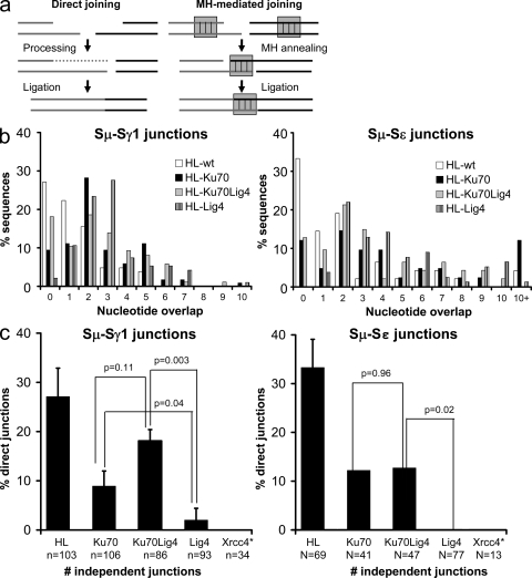
The classical nonhomologous end-joining (C-NHEJ) DNA double-strand break (DSB) repair pathway employs the Ku70/80 complex (Ku) for DSB recognition and the XRCC4/DNA ligase 4 (Lig4) complex for ligation. During IgH class switch recombination (CSR) in B lymphocytes, switch (S) region DSBs are joined by C-NHEJ to form junctions either with short microhomologies (MHs; "MH-mediated" joins) or no homologies ("direct" joins). In the absence of XRCC4 or Lig4, substantial CSR occurs via "alternative" end-joining (A-EJ) that generates largely MH-mediated joins. Because upstream C-NHEJ components remain in XRCC4- or Lig4-deficient B cells, residual CSR might be catalyzed by C-NHEJ using a different ligase. To address this, we have assayed for CSR in B cells deficient for Ku70, Ku80, or both Ku70 and Lig4. Ku70- or Ku80-deficient B cells have reduced, but still substantial, CSR. Strikingly, B cells deficient for both Ku plus Lig4 undergo CSR similarly to Ku-deficient B cells, firmly demonstrating that an A-EJ pathway distinct from C-NHEJ can catalyze CSR end-joining. Ku-deficient or Ku- plus Lig4-deficient B cells are also biased toward MH-mediated CSR joins; but, in contrast to XRCC4- or Lig4-deficient B cells, generate substantial numbers of direct CSR joins. Our findings suggest that more than one form of A-EJ can function in CSR.


Figures

Figure 1.
Figure 1.
Ku70−/−HL and Ku70−/−Lig4−/−HL mice posses mature splenic B cells which proliferate in response to stimulation by αCD40/IL-4. (a) Disruption of the Ku70 and Lig4 genomic loci and absence of Ku70 and Lig4 proteins in Ku70−/−Lig4−/− mice. (left) Southern blotting: BamHI-digested genomic DNA was probed with Ku70- or Lig4-specific probes. (right) Thymus protein extracts were assayed for the presence of Ku70 or Lig4 proteins. K, kidney protein extracts; BM, bone marrow protein extracts. Both the Southern and the Western blotting were repeated two times on independent samples. (b) WT-HL, Ku70−/−HL and Ku70−/−Lig4−/−HL mice have mature splenic B cells. Whole cell suspensions from spleens of WT-HL, Ku70−/−HL, and Ku70−/−Lig4−/−HL were used to quantify the percentage of B220+IgM+ mature B cells. Data shown are representative of more than 10 experiments, with at least one mouse of each genotype per experiment. (c) Ku70−/−HL mature B cells proliferate at levels comparable to WT-HL after αCD40/IL-4 stimulation. Splenic B cells were enriched with B220 magnetic beads and counted by Trypan blue staining starting at day 1.5 from the beginning of the culture, to allow for the disappearance of non–B cells. Data shown are based on two independent stimulations and a total of 4 WT-HL and 5 Ku70−/−HL mice were used. Error bars are based on the relative cell number for each mouse.
Figure 2.
Figure 2.
High IgG1 and IgE CSR levels in Ku70−/−, Ku70−/−Lig4−/−, Ku80−/−, and Lig4−/− B cells. (a) Representative FACS panel for IgG1 CSR in B cells stimulated with αCD40 plus IL-4 and assayed at day 3.5. Data shown are representative of >10 experiments, with one or more mouse of each genotype per experiment. Table S1 contains a summary of all FACS experiments. (b) ELISA quantification of IgM, IgG1, and IgE secretion (μG/ml) in B cells stimulated as in a. Black circles, H/H (the knock-in prerearranged heavy chain receptor is present on both alleles); white circles, H/+ (knock-in heavy chain on one allele). All experiments completed are shown, with one mouse per genotype per experiment and each circle represents one mouse. Table S2 contains a detailed summary of all ELISA experiments. (c) Frequency of IgG1-secreting hybridomas generated from αCD40/IL-4–stimulated B cells. Hybridomas for one mouse per genotype are shown. The number and percentage of hybridomas is indicated for each isotype assayed. “Other” denotes hybridomas negative for both IgM and IgG1 secretion, some of which may be IgE+ because IgE ELISA was not performed for the fusions shown. Data shown are representative of three independent hybridoma fusion experiments (one mouse each) for WT-HL and Ku70−/−HL and two independent hybridoma fusions (one mouse each) for the Ku70−/−Lig4−/−HL mice. Table S3 contains a summary of all hybridoma data, with multiple mice of each genotype.
Figure 3.
Figure 3.
Ku70- and Ku70/Lig4-deficient cells undergo robust CSR to IgG3 and IgG2b. (a) Representative FACS panel for IgG3 CSR in B cells stimulated with LPS and anti–IgD-dextran and assayed at day 3.5. Data shown are representative of >10 experiments, with one or more mouse of each genotype per experiment. Table S1 contains a summary of all FACS experiments. (b) ELISA quantification of IgM, IgG3, and IgG2b secretion (micrograms/milliliter) in B cells stimulated as in a. Black circles, H/H (the knock-in prerearranged heavy chain receptor is present on both alleles); white circles: H/+ (knock-in heavy chain on one allele). Each circle represents one mouse. All experiments completed are shown, with one mouse per genotype per experiment. Table S2 contains a summary of all ELISA experiments. (c) Representative ELISPOT assay on B cells stimulated as in A. (top) Dots represent Igκ- and Igλ-secreting B cells. (bottom) Dots represent IgG3-secreting B cells. Percentages of B cells undergoing CSR to IgG3 are shown. Data shown are representative of three experiments, with one mouse of each genotype per experiment.
Figure 4.
Figure 4.
Time course for IgG1 CSR in Ku70−/−H/HL+, Ku70−/−Lig4−/−H/HL+, and WT-H/HL+ B cells stimulated with αCD40 plus IL-4. (a) Representative FACS plots for IgG1 CSR quantification in B cells from the indicated backgrounds stimulated with αCD40 plus IL-4. IgG1 FACS was recorded at day 2.5, 3.5, and 4.5. AID, AID−/− B cells used as negative controls for CSR. Percentage of B220+IgG1+ cells that have undergone CSR is shown for each genotype at each time point. Data shown are representative of three independent B cell stimulation experiments, with one mouse of each genotype per experiment. (b) Quantification of CSR levels at days 2.5, 3.5, and 4.5 from the beginning of the in vitro stimulation. Data are based on at least three mice per genotype used in three separate experiments. Table S1 contains the summary of all FACS experiments.
Figure 5.
Figure 5.
The Ku-dependent A-EJ pathway uses a distinct mechanism for S region joining. (a) Classical NHEJ performs both direct (left) and MH-mediated (right) end-joining. Direct joining occurs when DNA ends are joined either immediately, or after processing (removal of overhangs or fill-in by polymerases). MH-mediated joining (right) is facilitated by base-pairing interactions between short stretches of nucleotides at or near the ends of the DSB. (b) MH length in Sμ-Sγ1 (left) and Sμ-Sε (right) junctions from Ku70−/−HL, Ku70−/−Lig4−/−HL, Lig4−/−HL and WT/HL B cells stimulated in culture with αCD40 plus IL-4. Ku70-, Ku70/Lig4-, and Lig4-deficient cells have more Sμ-Sγ1 (left) and Sμ-Sε (right) joins with longer MH than WT. Data shown are based on 86 or more independent sequences per genotype for Sμ-Sγ1 junctions and 41 or more independent sequences for Sμ-Sε junctions. At least three mice per genotype in three independent experiments were used for Sμ-Sγ1 junctions and one Ku70−/−HL mouse, two Ku70−/−Lig4−/−HL mice, three Lig4−/−HL, and three WT-HL mice in three independent experiments were used for Sμ-Sε junctions. A detailed summary of all S region junction isolation experiments and mice is presented in Table S4. (C) Percentage of direct Sμ-Sγ1 (left) and Sμ-Sε (right) joins in Ku70−/−HL, Ku70−/−Lig4−/−HL, Lig4−/−HL, XRCC4−/−HL and WT/HL B cells. Ku70 and Ku70Lig4-deficient cells have significantly more direct Sμ-Sγ1 and Sμ-Sε joins than Lig4 germline-deficient cells. *, XRCC4 junctions were acquired and published previously (Yan et al., 2007) and are shown here for comparison. Two-tailed Student’s t test was used for p-value. Data shown are based on 86 or more independent sequences per genotype for Sμ-Sγ1 junctions and 41 or more independent sequences for Sμ-Sε junctions. At least three mice per genotype in three independent experiments were used for Sμ-Sγ1 junctions, and one Ku70−/−HL mouse, two Ku70−/−Lig4−/−HL mice, three Lig4−/−HL, and three WT-HL mice in three independent experiments were used for Sμ-Sε junctions. N, number of independent sequences.

Similar articles

Cited by

References

    1. Adachi N., Ishino T., Ishii Y., Takeda S., Koyama H. 2001. DNA ligase IV-deficient cells are more resistant to ionizing radiation in the absence of Ku70: implications for DNA double-strand break repair. Proc. Natl. Acad. Sci. USA. 98:12109–12113 10.1073/pnas.201271098 - DOI - PMC - PubMed
    1. Audebert M., Salles B., Calsou P. 2004. Involvement of poly(ADP-ribose) polymerase-1 and XRCC1/DNA ligase III in an alternative route for DNA double-strand breaks rejoining. J. Biol. Chem. 279:55117–55126 10.1074/jbc.M404524200 - DOI - PubMed
    1. Bassing C.H., Alt F.W. 2004. The cellular response to general and programmed DNA double strand breaks. DNA Repair (Amst.). 3:781–796 10.1016/j.dnarep.2004.06.001 - DOI - PubMed
    1. Boulton S.J., Jackson S.P. 1996. Saccharomyces cerevisiae Ku70 potentiates illegitimate DNA double-strand break repair and serves as a barrier to error-prone DNA repair pathways. EMBO J. 15:5093–5103 - PMC - PubMed
    1. Callén E., Jankovic M., Wong N., Zha S., Chen H.T., Difilippantonio S., Di Virgilio M., Heidkamp G., Alt F.W., Nussenzweig A., Nussenzweig M. 2009. Essential role for DNA-PKcs in DNA double-strand break repair and apoptosis in ATM-deficient lymphocytes. Mol. Cell. 34:285–297 10.1016/j.molcel.2009.04.025 - DOI - PMC - PubMed

Publication types