. 2010 Mar 16;107(11):5214-9.
doi: 10.1073/pnas.0910772107. Epub 2010 Feb 8.

A developmental framework for endodermal differentiation and polarity

Affiliations

A developmental framework for endodermal differentiation and polarity

Julien Alassimone et al. Proc Natl Acad Sci U S A. .

Abstract

The endodermis is a root cell layer common to higher plants and of fundamental importance for root function and nutrient uptake. The endodermis separates outer (peripheral) from inner (central) cell layers by virtue of its Casparian strips, precisely aligned bands of specialized wall material. Here we reveal that the membrane at the Casparian strip is a diffusional barrier between the central and peripheral regions of the plasma membrane and that it mediates attachment to the extracellular matrix. This membrane region thus functions like a tight junction in animal epithelia, although plants lack the molecular modules that establish tight junction in animals. We have also identified a pair of influx and efflux transporters that mark both central and peripheral domains of the plasma membrane. These transporters show opposite polar distributions already in meristems, but their localization becomes refined and restricted upon differentiation. This "central-peripheral" polarity coexists with the apical-basal polarity defined by PIN proteins within the same cells, but utilizes different polarity determinants. Central-peripheral polarity can be already observed in early embryogenesis, where it reveals a cellular polarity within the quiescent center precursor cell. A strict diffusion block between polar domains is common in animals, but had never been described in plants. Yet, its relevance to endodermal function is evident, as central and peripheral membranes of the endodermis face fundamentally different root compartments. Further analysis of endodermal transporter polarity and manipulation of its barrier function will greatly promote our understanding of plant nutrition and stress tolerance in roots.


Conflict of interest statement

The authors declare no conflict of interest.

Figures
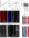
Fig. 1.
Fig. 1.
Molecular and quantitative analysis of endodermal differentiation. (A) Average rate and duration of endodermal cell elongation in 9-h time-series/10-min intervals (13 cells/6 roots). (B) Average cell length vs. position in the cell file (n = 25). Red arrows mark the position of the differentiation events depicted in D–G. (C) Penetration of propidium iodide (PI) into the stele (Left) is blocked in differentiated roots (Right). Block at 14.4 cells (n = 30) is shown. (D) Casparian strip presence visualized by autofluorescence is observed at 11.7 cells (n = 25). (E) Cell-wall adhesion of the CSD membrane upon plasmolysis. Attachment to transversal walls is observed at 11.9 cells (n = 21) (Right). Before that, protoplasts retract from each other (Left). (F) The membrane tracer FM4-64 highlights all surfaces of endodermal and inner cells (Left), but becomes restricted to the outer domain of the endodermis in differentiated roots (Right) at 11.4 cells (n = 30). (G) Exclusion of PM marker NPSN12 from the Casparian strip domain (CSD), observed at 10.9 cells (n = 31). Left and Upper Right are before differentiation, and Middle and Lower Right are after differentiation. (H) Graph of the positioning of the different differentiation events, as shown in C–G. Red lines mark the narrow zone (∼11–13 cells) in which differentiation events occur. Propidium iodide is probably observed later due to some upward diffusion or mass flow from undifferentiated tissue. (I) Cartoon depicting the compartments of the endodermis: inner apoplastic space of vasculature (black), outer apoplastic space of cortex (red), Casparian strip blocking extracellular diffusion (blue), PMs (white), PM region (CSD) mediating cell wall attachment, and protein exclusion and suppression of FM4-64 (black dots). 3D representation in the same color code visualizes how two “dots” in median longitudinal sections in C–G relate to ring structures of the CS in three dimensions. en, endodermis; ct, cortex; st, stele. Arrowheads in C–G indicate position of the CS/CSD, and in E they indicate the presumptive transversal borders between cells. (Scale bars: C–G, 10 μm; G Right, 5 μm.)
Fig. 2.
Fig. 2.
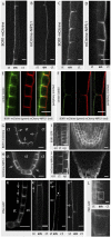
Endodermal polarity is different from PIN polarity and organizes with respect to the stele. (AD) Signals of BOR-mCit and mCit-NIP5;1 at opposite cell sides in differentiated (A and B) and meristematic (C and D) cells. (E and F) Colocalization of BOR1-mCit (Left), mCherry-NIP5;1 (Center), and overlay (Right). Colocalization can be observed in meristematic (E), but not in differentiated cells (F). (G and H) Localization of BOR1-mCit and mCit-NIP5;1 in quiescent center and initials and proendodermal cells. BOR1 signals polarize toward NIP5;1 signals away from the stele. (I and J) Signals of BOR1 and NIP5;1 in nonendodermal cells, expressed from 35S or UBQ10 promoter, respectively. Signals from neighboring cells are confounded, and the same orientation of polarity is observed as in the endodermis. (K) Localization of PIN2-GFP is apolar in quiescent center and basal in initials and proendodermal cells. Localization switches to apical in elongating cells (K′) and to the central side in differentiated cells (K′′ and K′′′). (L) PIN1-GFP shows basal localization in endodermis. Overview (Upper) and zoom in (Lower) are shown. ct, cortex; en, endodermis; ep, epidermis; st, stele. Open arrows indicate direction of polarity of individual cells. Arrowhead indicates position of the CSD (Scale bar: 10 μm.)
Fig. 3.
Fig. 3.
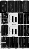
Central–peripheral polarity becomes established early in embryogenesis. (A–C) BOR1-mCit signals in quiescent center precursor cells, initials, and proendodermal cells display polarity oriented toward the center of the embryo. Arrows point to cell sides with visible polar accumulation. (Left) Transmitted light image. (Right) Confocal image. (D) Number of embryos observed with polar BOR1-mCit signals over total number of embryos inspected. (E–G) mCit-NIP5;1 signals in quiescent center precursor cells, initials, and proendodermal cells display polarity oriented toward the periphery of the embryo. Arrows point to cell sides with visible polar accumulation. (Left) Transmitted light image. (Right) Confocal image. (H) Number of embryos observed with polar mCit-NIP5;1 signals over total number of embryos inspected. (Scale bar: 10 μm.)
Fig. 4.
Fig. 4.
Dependence of endodermal differentiation and polarity on actin and vesicle trafficking. (A) 35S::YFP-Fimbrin highlights actin during root hair formation in epidermis. (B and C) Surface (B) and median optical section (C) of SCR::YFP-Fimbrin. No localized accumulation of actin can be seen. (D) Signals after 1 h treatment with 10 μM LatB. (E, G, and H) Localization of BOR1, NIP5;1, or NPSN12 before and after differentiation (top and bottom) at the PM is unaffected after LatB treatment (10 μM, 1 h). (F) No shift in meristem distance is observed for the NPSN12 depletion zone after LatB treatment (10 μM, 5 h). (I) PM localization of NPSN12 is unaffected after BFA treatment (50 μM, 90 min) before (Left) and after (Right) differentiation. (J) No shift in meristem distance for the NPSN12 depletion zone is observed after BFA treatment (50 μM, 5 h). (K and L) BOR1 and NIP5;1 polarity is unaffected by BFA treatment (50 μM, 90 min). (M) WM treatment (50 μM, 1 h) effects of YFP-RabF2b, a WM-sensitive compartment marker in endodermis and stele, as seen by the higher background, irregular dots, and ring-like structures (Lower), compared to control (Upper). (N) WM treatment (50 μM, 1 h) leaves the NPSN12 depletion zone (CSD) intact (Left) and does not affect localization before differentiation (Left). (O) WM treatment (50 μM, 1 h) does not affect BOR1 polarity in elongating cells. (P) WM treatment (50 μM, 1 h) leads to complete depolarization of NIP5;1 in cells before differentiation (Left), but does not affect polarity in differentiated cells (Right). Arrowheads indicate position of the CSD. (Scale bars: 10 μm and (E) 5 μm.)

References

    1. Enstone DE, Peterson CA, Ma F. Root endodermis and exodermis: Structure, function, and responses to the environment. J Plant Growth Regul. 2002;21:335–351.
    1. Cui H, et al. An evolutionarily conserved mechanism delimiting SHR movement defines a single layer of endodermis in plants. Science. 2007;316:421–425. - PubMed
    1. Zeier J, Schreiber L. Chemical composition of hypodermal and endodermal cell walls and xylem vessels isolated from Clivia miniata (identification of the biopolymers lignin and suberin) Plant Physiol. 1997;113:1223–1231. - PMC - PubMed
    1. Bonnett HT., Jr The root endodermis: Fine structure and function. J Cell Biol. 1968;37:199–205. - PMC - PubMed
    1. Ma JF, et al. An efflux transporter of silicon in rice. Nature. 2007;448:209–212. - PubMed

Publication types