. 2010 Feb 23;107(8):3463-8.
doi: 10.1073/pnas.0913083107. Epub 2010 Feb 8.

Histidine-mediated RNA transfer to GDP for unique mRNA capping by vesicular stomatitis virus RNA polymerase

Affiliations

Histidine-mediated RNA transfer to GDP for unique mRNA capping by vesicular stomatitis virus RNA polymerase

Tomoaki Ogino et al. Proc Natl Acad Sci U S A. .

Abstract

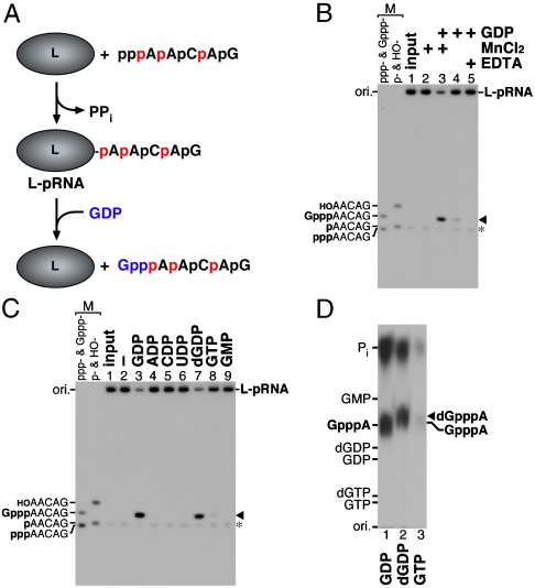
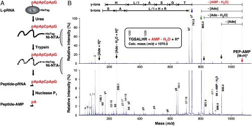
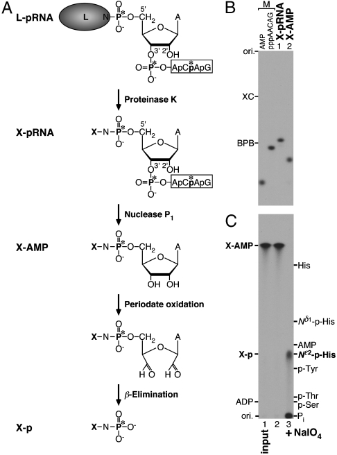
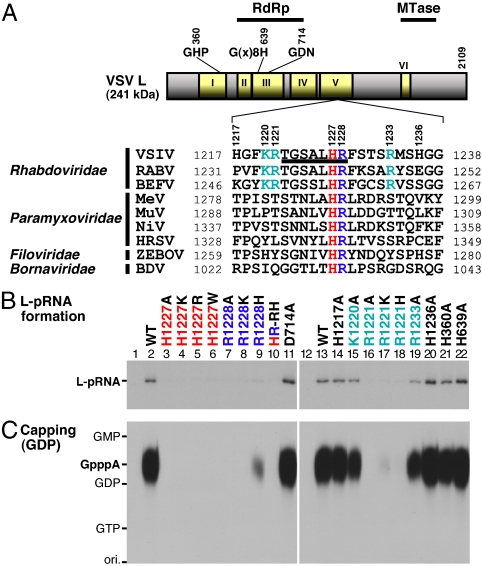
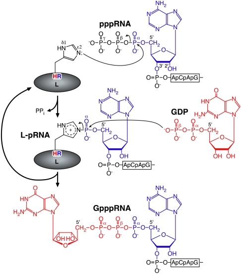
The RNA-dependent RNA polymerase L protein of vesicular stomatitis virus, a prototype of nonsegmented negative-strand (NNS) RNA viruses, forms a covalent complex with a 5'-phosphorylated viral mRNA-start sequence (L-pRNA), a putative intermediate in the unconventional mRNA capping reaction catalyzed by the RNA:GDP polyribonucleotidyltransferase (PRNTase) activity. Here, we directly demonstrate that the purified L-pRNA complex transfers pRNA to GDP to produce the capped RNA (Gpp-pRNA), indicating that the complex is a bona fide intermediate in the RNA transfer reaction. To locate the active site of the PRNTase domain in the L protein, the covalent RNA attachment site was mapped. We found that the 5'-monophosphate end of the RNA is linked to the histidine residue at position 1,227 (H1227) of the L protein through a phosphoamide bond. Interestingly, H1227 is part of the histidine-arginine (HR) motif, which is conserved within the L proteins of the NNS RNA viruses including rabies, measles, Ebola, and Borna disease viruses. Mutagenesis analyses revealed that the HR motif is required for the PRNTase activity at the step of the enzyme-pRNA intermediate formation. Thus, our findings suggest that an ancient NNS RNA viral polymerase has acquired the PRNTase domain independently of the eukaryotic mRNA capping enzyme during evolution and PRNTase becomes a rational target for designing antiviral agents.


Conflict of interest statement

The authors declare no conflict of interest.

Figures

Fig. 1.
Fig. 1.
RNA transfer from the purified L-pRNA complex to GDP. (A) The VSV L protein was incubated with pppAACAG labeled with [α-32P]AMP (32P shown in red) to generate the covalent L-pRNA complex. The purified L-pRNA complex was incubated with GDP (blue) to examine whether pRNA linked to the L protein is transferred to GDP to form the capped RNA. (B) The purified L-pRNA complex was incubated with GDP in the presence or absence of MnCl2 or EDTA, as indicated. The reaction mixtures were analyzed by urea-20% PAGE followed by autoradiography. Marker (M) lanes indicate the AACAG RNAs with different 5′-ends (ppp-, triphosphorylated; Gppp-, capped; p-, monophosphorylated; HO-, hydroxyl). Lane 1 indicates the input L-pRNA complex. The arrowhead and asterisk indicate the capped RNA (Gpp-pAACAG) and free pRNA, respectively. (C and D) The purified L-pRNA complex was incubated with the indicated nucleotide in the presence of MnCl2. The reaction mixtures were analyzed by urea-20% PAGE (C). RNAs released with GDP (lane 3), dGDP (lane 7), and GTP (lane 8) (indicated by the arrowhead) were purified from the gel and digested with nuclease P1 and CIAP. The digests were analyzed along with marker compounds by PEI-cellulose TLC (D). The positions of standard marker compounds, visualized under UV light at wavelength of 254 nm, are shown on the left. The position of the putative 2′-deoxyguanosine(5′)triphospho(5′)adenosine (dGpppA) cap structure is shown by the arrowhead.
Fig. 2.
Fig. 2.
Localization of the covalent RNA attachment site in the L protein. (A) The His-tagged L-pRNA complex (pRNA shown in red) was denatured with urea and purified with Ni-NTA agarose. The denatured complex on the resin was digested with trypsin. The resulting peptide-pRNA complex was digested with nuclease P1 to generate the peptide-AMP complex. (B) The purified peptide-AMP complex was analyzed by MALDI-TOF MS (see Fig. S4) and its singly charged ion at m/z 1070.6 was subjected to MS/MS analysis. MS/MS spectra are given as differently expanded intensity axes (Upper and Lower panels). The parent ion ([M + H]+) is shown by the red arrow. Other vertical arrows indicate putative fragment ions that presumably lost adenine (Ade) (C5H5N5, 135.1 Da), anhydro-adenosine (Ado) (C10H11N5O3, 249.1 Da), Ado (C10H13N5O4, 267.1 Da), and anhydro-AMP (C10H12N5O6P1, 329.1 Da) from the parent ion and protonated Ade and anhydro-Ado ions ([Ade + H]+ and [Ado - H2O + H]+, respectively). The asterisk indicates the putative b6 ion linked to AMP (b6 + AMP - H2O). Deduced peptide fragment ions (b- and y-ions) lacking the AMP moiety and immonium ions are indicated above the signals. The horizontal arrows indicate an amino acid sequence [TGSA(L/I)HR] elucidated from the b- and y-ion series. The sequence corresponds to residues 1,222 to 1,228 of the VSV L protein. The calc. monoisotopic mass of the peptide-AMP complex ion is shown.
Fig. 3.
Fig. 3.
Analysis of the polyribonucleotidylated amino acid in the L protein. (A) The L-pRNA complex (asterisks indicate 32P) was digested with proteinase K to obtain a single amino acid (X)-pRNA complex. After removal of the RNA chain from the X-pRNA complex by nuclease P1 digestion, the resulting X-AMP complex was subjected to periodate oxidation followed by β-elimination to generate phosphoamino acid (X-p). (B) The X-pRNA and X-AMP complexes were analyzed by urea-20% PAGE followed by autoradiography. (C) The X-AMP complex was incubated with (lane 3) or without (lane 2) sodium periodate in an alkaline solution (pH 10.5) at room temperature. After adding an excess amount of ethylene glycol, the reaction mixtures were incubated at 50 °C. The reaction mixtures were analyzed by reverse phase silica gel TLC with marker compounds including phosphoserine (p-Ser), phosphothreonine (p-Thr), phosphotyrosine (p-Tyr), Nδ1-phosphohistidine (Nδ1-p-His), Nε2-phosphohistidine (Nε2-p-His), histidine (His), AMP, and ADP. Lane 1 indicates the input X-AMP complex.
Fig. 4.
Fig. 4.
The HR motif in the L protein is required for the PRNTase activity. (A) A schematic structure of the VSV L protein is shown with six amino acid sequence blocks (I–VI) conserved in the NNS RNA viral L proteins. The positions of the putative RNA-dependent RNA polymerase (RdRp) and cap methyltransferase (MTase) domains are indicated on the top. The local sequence containing the covalent RNA attachment site (H1227) of the VSV L protein is aligned with those of representative NNS RNA viral L proteins (see Fig. S5). The sequence of the tryptic peptide linked to the RNA (Fig. 2) is underlined. The conserved H and R residues are shown in red and blue, respectively. Basic amino acid residues conserved only in the L proteins of rhabdoviruses are shown in cyan. VSIV, vesicular stomatitis Indiana virus; RABV, rabies virus; BEFV, bovine ephemeral fever virus; MeV, measles virus; MuV, mumps virus; NiV, Nipah virus; HRSV, human respiratory syncytial virus; ZEBOV, Zaire ebolavirus; BDV, Borna disease virus. (B) The WT and mutant L proteins (see Fig. S6A) were incubated with 32P-labeled pppAACAG. The resulting L-pRNA complex was analyzed by SDS-7.5% PAGE followed by autoradiography. Lanes 1 and 12 indicate no L protein. (C) The WT and mutant L proteins (see panel B) were subjected to the capping reactions with pppAACAG and [α-32P]GDP as substrates. CIAP and nuclease P1-resistant products were analyzed by PEI-cellulose TLC followed by autoradiography. Lanes 1 and 12 indicate no L protein. The positions of standard marker compounds are shown on the left.
Fig. 5.
Fig. 5.
A proposed model of the polyribonucleotidyl transfer reaction catalyzed by the unconventional mRNA capping enzyme L protein. For detail, see text.

Comment in

  • Viruses know more than one way to don a cap.
    Koonin EV, Moss B. Koonin EV, et al. Proc Natl Acad Sci U S A. 2010 Feb 23;107(8):3283-4. doi: 10.1073/pnas.0915061107. Epub 2010 Feb 18. Proc Natl Acad Sci U S A. 2010. PMID: 20167804 Free PMC article. No abstract available.

Similar articles

Cited by

References

    1. Banerjee AK. 5′-terminal cap structure in eucaryotic messenger ribonucleic acids. Microbiol Rev. 1980;44:175–205. - PMC - PubMed
    1. Furuichi Y, Shatkin AJ. Viral and cellular mRNA capping: Past and prospects. Adv Virus Res. 2000;55:135–184. - PMC - PubMed
    1. Shuman S. Structure, mechanism, and evolution of the mRNA capping apparatus. Prog Nucleic Acid Res Mol Biol. 2001;66:1–40. - PubMed
    1. Ogino T, Banerjee AK. Unconventional mechanism of mRNA capping by the RNA-dependent RNA polymerase of vesicular stomatitis virus. Mol Cell. 2007;25:85–97. - PubMed
    1. Ogino T, Banerjee AK. Formation of guanosine(5′)tetraphospho(5′)adenosine cap structure by an unconventional mRNA capping enzyme of vesicular stomatitis virus. J Virol. 2008;82:7729–7734. - PMC - PubMed

Publication types

MeSH terms

LinkOut - more resources