. 2010 Mar 2;107(9):4051-6.
doi: 10.1073/pnas.0911060107. Epub 2010 Feb 8.

Stem cell-specific activation of an ancestral myc protooncogene with conserved basic functions in the early metazoan Hydra

Affiliations

Stem cell-specific activation of an ancestral myc protooncogene with conserved basic functions in the early metazoan Hydra

Markus Hartl et al. Proc Natl Acad Sci U S A. .

Abstract

The c-myc protooncogene encodes a transcription factor (Myc) with oncogenic potential. Myc and its dimerization partner Max are bHLH-Zip DNA binding proteins controlling fundamental cellular processes. Deregulation of c-myc leads to tumorigenesis and is a hallmark of many human cancers. We have identified and extensively characterized ancestral forms of myc and max genes from the early diploblastic cnidarian Hydra, the most primitive metazoan organism employed so far for the structural, functional, and evolutionary analysis of these genes. Hydra myc is specifically activated in all stem cells and nematoblast nests which represent the rapidly proliferating cell types of the interstitial stem cell system and in proliferating gland cells. In terminally differentiated nerve cells, nematocytes, or epithelial cells, myc expression is not detectable by in situ hybridization. Hydra max exhibits a similar expression pattern in interstitial cell clusters. The ancestral Hydra Myc and Max proteins display the principal design of their vertebrate derivatives, with the highest degree of sequence identities confined to the bHLH-Zip domains. Furthermore, the 314-amino acid Hydra Myc protein contains basic forms of the essential Myc boxes I through III. A recombinant Hydra Myc/Max complex binds to the consensus DNA sequence CACGTG with high affinity. Hybrid proteins composed of segments from the retroviral v-Myc oncoprotein and the Hydra Myc protein display oncogenic potential in cell transformation assays. Our results suggest that the principal functions of the Myc master regulator arose very early in metazoan evolution, allowing their dissection in a simple model organism showing regenerative ability but no senescence.


Conflict of interest statement

The authors declare no conflict of interest.

Figures

Fig. 1.
Fig. 1.
Amino acid sequences of Hydra Myc1 and Max proteins and alignment with their vertebrate homologs. (A) Alignment of human (Hu) c-Myc, chicken (Ck) c-Myc, and Hydra (Hy) Myc1 sequences (GenBank accession nos.: hu c-Myc, NP_002458; ck c-Myc, NP_001026123; hy Myc1, GQ856263). (B) Alignment of human, chicken, and Hydra Max sequences (GenBank accession nos.: hu Max, NP_002373; ck Max, P52162; hy Max, GQ856264). Identical residues are shaded in Blue, gaps are indicated by Dashes. The positions of the Myc boxes (MB) I–IV in the transactivation domains of the vertebrate proteins and of the bHLH-Zip region in the Myc or Max DNA binding domains are indicated. Conserved heptad repeat residues in the leucine zipper regions are marked by Asterisks. The positions corresponding to exon-exon junctions are indicated by Arrows above (HU, CK) and below (HY) the alignments. Alignments were generated by using the ClustalW algorithm with additional manual adjustments.
Fig. 2.
Fig. 2.
Expression of Hydra myc1 and max genes and their protein products. (A) Northern analyses using aliquots (2.0 μg) of poly(A)+-selected RNAs from whole Hydra animals and Hydra myc1 or max specific cDNA probes. Each filter was stripped and rehybridized with a Hydra CAD specific cDNA probe. (B) Immunoprecipitation analysis using aliquots (5 × 106 cpm) of boiled cell extracts from [35S]methionine-labeled Hydra animals and a polyclonal antiserum directed against Hydra Myc1 recombinant protein (α-hy Myc1), or normal rabbit serum (NRS). For comparison, [35S]methionine-labeled Hydra Myc1 p39 and p36 proteins were also produced by in vitro translation of corresponding cDNAs cloned in Bluescript (BS) vectors and immunoprecipitated. Proteins were analyzed by SDS/PAGE (10%, wt/vol). (C) Immunoblot analysis of a 30-μg aliquot of total cell proteins from whole Hydra polyps and of in vitro translated Hydra Max using a polyclonal antiserum directed against Hydra Max recombinant protein (α-hy Max). Proteins were resolved by SDS/PAGE (12.5%, wt/vol).
Fig. 3.
Fig. 3.
Expression patterns of Hydra myc1 and max visualized by in situ hybridization. (A, B) myc1 and max are activated in cells belonging to the interstitial stem cell lineage in the gastric region of intact, budding polyps. In addition, max is expressed at a lower level in the epithelium throughout the entire body column. (CF) myc1 expression in interstitial stem cells (C and E), proliferating nematoblast nests (D and F), and a gland cell (Insert in C and E), visualized by in situ hybridization in macerated single cell preparations. (C and D) bright field optics; (E and F) phase contrast optics. Ecto: ectodermal epithelial cell; Endo: endodermal epithelial cell; and NV: nerve cell. (G) Scheme of the differentiation pathways in the interstitial stem cell system with myc1 expressing subpopulations shaded in Yellow.
Fig. 4.
Fig. 4.
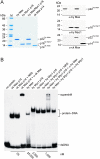
Hydra Myc1 and Max recombinant proteins and their biochemical activity. (A) SDS/PAGE (5.0–17.5% gradient, wt/vol) of 2-μg (Coomassie brilliant blue staining) or 50-ng (immunoblotting) aliquots of purified recombinant Hydra Myc1 p16 (amino acids 201–314), Hydra Max, a 1∶1 mixture of both proteins, and chicken Max p14 (amino acids 22–113). Specific antibodies are indicated below the Blots. (B) EMSA using the recombinant proteins shown in A and 0.3-ng (25,000 cpm) aliquots of a [32P]-labeled double-stranded 18-mer deoxyoligonucleotide containing the Myc/Max-binding motif 5′-CACGTG-3′. Antibodies were added to the binding reactions as indicated. Final protein concentrations are indicated below.
Fig. 5.
Fig. 5.
Cell transforming activities of Hydra and viral Myc hybrid proteins. (A) Schematic diagram of the coding regions of v-myc (Blue), and of hy/v-myc and v/hy-myc hybrids (Hydra sequences shown in Yellow). The constructs were inserted into the unique ClaI site of the replication-competent retroviral pRCAS vector used for DNA transfection into quail embryo fibroblasts (QEF). (B) Immunoprecipitation of endogenous c-Myc and ectopic Myc proteins using 1.0 × 107-cpm aliquots of lysates from [35S]methionine-labeled QEFs transfected with the pRCAS constructs shown in A, and antibodies directed against amino-terminal (N) or carboxyl-terminal (C) segments of v-Myc, or normal rabbit serum (NRS). Proteins were resolved by SDS/PAGE (10%, wt/vol). (C) Proliferation rates of QEFs transfected with the pRCAS constructs shown in A and passaged several times. Equal numbers (7.5 × 105) of cells were seeded onto 60-mm dishes, and cell numbers were determined at the indicated time points. (D) Top: QEFs on 60-mm dishes were transfected with 4-μg aliquots of DNA from the pRCAS constructs shown in A, kept under agar overlay for 2 wk, and then stained with eosin methylene blue. Numbers of foci per dish are indicated. Middle: QEFs were transfected with the pRCAS constructs shown in A and passaged several times. The doubling times (Td) of the cell populations are indicated below the phase-contrast micrographs. Bottom: Equal numbers (1.0 × 105) of transfected and passaged cells were seeded in soft agar and incubated for 2 wk. Numbers of colonies per 1,000 cells seeded are shown below the bright-field micrographs.

Similar articles

Cited by

References

    1. Bister K, Jansen HW. Oncogenes in retroviruses and cells: biochemistry and molecular genetics. Adv Cancer Res. 1986;47:99–188. - PubMed
    1. Eisenman RN. Deconstructing Myc. Genes Dev. 2001;15:2023–2030. - PubMed
    1. Eilers M, Eisenman RN. Myc’s broad reach. Genes Dev. 2008;22:2755–2766. - PMC - PubMed
    1. Nesbit CE, Tersak JM, Prochownik EV. MYC oncogenes and human neoplastic disease. Oncogene. 1999;18:3004–3016. - PubMed
    1. Dang CV, Kim JW, Gao P, Yustein J. The interplay between MYC and HIF in cancer. Nat Rev Cancer. 2008;8:51–56. - PubMed

Publication types

MeSH terms

Substances

Associated data