

Brain perivascular macrophages and the sympathetic response to inflammation in rats after myocardial infarction
- PMID: 20142564
- PMCID: PMC2890291
- DOI: 10.1161/HYPERTENSIONAHA.109.142836
Brain perivascular macrophages and the sympathetic response to inflammation in rats after myocardial infarction
Abstract
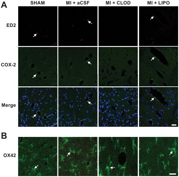
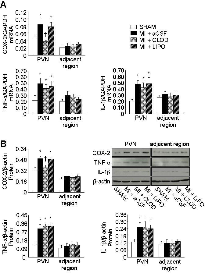
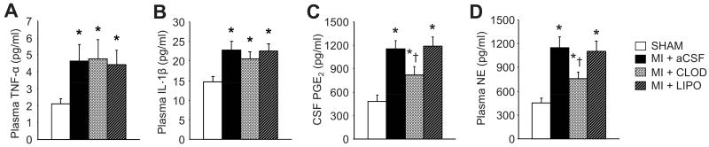
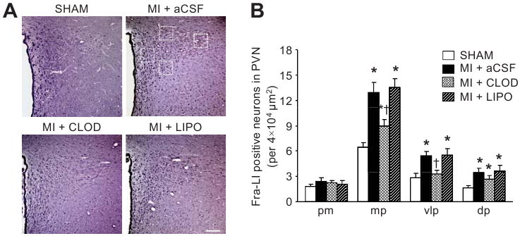
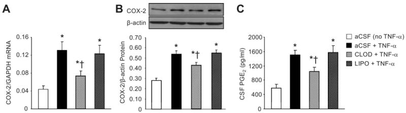
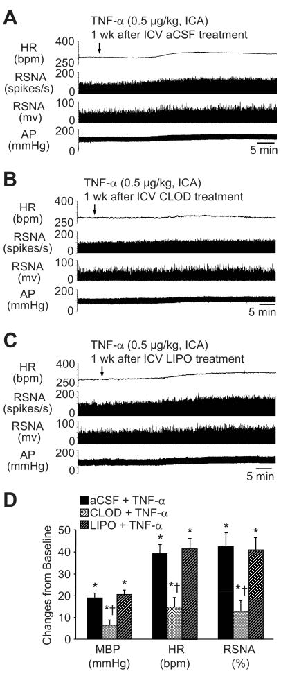
Inflammation is associated with increased sympathetic drive in cardiovascular diseases. Blood-borne proinflammatory cytokines, markers of inflammation, induce cyclooxygenase 2 (COX-2) activity in perivascular macrophages of the blood-brain barrier. COX-2 generates prostaglandin E(2), which may enter the brain and increase sympathetic nerve activity. We examined the contribution of this mechanism to augmented sympathetic drive in rats after myocardial infarction (MI). Approximately 24 hours after acute MI, rats received an intracerebroventricular injection (1 microL/min over 40 minutes) of clodronate liposomes (MI+CLOD) to eliminate brain perivascular macrophages, liposomes alone, or artificial cerebrospinal fluid. A week later, COX-2 immunoreactivity in perivascular macrophages and COX-2 mRNA and protein had increased in hypothalamic paraventricular nucleus of MI rats treated with artificial cerebrospinal fluid or liposomes alone compared with sham-operated rats. In MI+CLOD rats, neither perivascular macrophages nor COX-2 immunoreactivity was seen in the paraventricular nucleus, and COX-2 mRNA and protein levels were similar to those in sham-operated rats. Prostaglandin E(2) in cerebrospinal fluid, paraventricular nucleus neuronal excitation, and plasma norepinephrine were less in MI+CLOD rats than in MI rats treated with artificial cerebrospinal fluid or liposomes alone but more than in sham-operated rats. Intracerebroventricular CLOD had no effect on interleukin 1beta and tumor necrosis factor-alpha mRNA and protein in the paraventricular nucleus or plasma interleukin-1beta and tumor necrosis factor-alpha, which were increased in MI compared with sham-operated rats. In normal rats, pretreatment with intracerebroventricular CLOD reduced (P<0.05) the renal sympathetic, blood pressure, and heart rate responses to intracarotid artery injection of tumor necrosis factor-alpha (0.5 microg/kg); intracerebroventricular liposomes had no effect. The results suggest that proinflammatory cytokines stimulate sympathetic excitation after MI by inducing COX-2 activity and prostaglandin E(2) production in perivascular macrophages of the blood-brain barrier.
Conflict of interest statement
Figures
Comment in
-
Brain perivascular macrophages and central sympathetic activation after myocardial infarction: heart and brain interaction.Hypertension. 2010 Mar;55(3):610-1. doi: 10.1161/HYPERTENSIONAHA.109.145128. Epub 2010 Feb 8. Hypertension. 2010. PMID: 20142562 No abstract available.
References
-
- Granger JP. An emerging role for inflammatory cytokines in hypertension. Am J Physiol Heart Circ Physiol. 2006;290:H923–H924. - PubMed
-
- Perin PC, Maule S, Quadri R. Sympathetic nervous system, diabetes, and hypertension. Clin Exp Hypertens. 2001;23:45–55. - PubMed
-
- Yu Y, Kang YM, Zhang ZH, Wei SG, Chu Y, Weiss RM, Felder RB. Increased cyclooxygenase-2 expression in hypothalamic paraventricular nucleus in rats with heart failure: role of nuclear factor kappaB. Hypertension. 2007;49:511–518. - PubMed
-
- Yu Y, Zhang ZH, Wei SG, Chu Y, Weiss RM, Heistad DD, Felder RB. Central gene transfer of interleukin-10 reduces hypothalamic inflammation and evidence of heart failure in rats after myocardial infarction. Circ Res. 2007;101:304–312. - PubMed
-
- Straznicky NE, Eikelis N, Lambert EA, Esler MD. Mediators of sympathetic activation in metabolic syndrome obesity. Curr Hypertens Rep. 2008;10:440–447. - PubMed
Publication types
MeSH terms
Substances
Grants and funding
LinkOut - more resources
Full Text Sources
Other Literature Sources
Medical
Research Materials
