Review
. 2010 Apr;87(4):E239-54.
doi: 10.1097/OPX.0b013e3181d39464.

Dynamic regulation of barrier integrity of the corneal endothelium

Affiliations
Review

Dynamic regulation of barrier integrity of the corneal endothelium

Sangly P Srinivas. Optom Vis Sci. 2010 Apr.

Abstract

The corneal endothelium maintains stromal deturgescence, which is a prerequisite for corneal transparency. The principal challenge to stromal deturgescence is the swelling pressure associated with the hydrophilic glycosaminoglycans in the stroma. This negative pressure induces fluid leak into the stroma from the anterior chamber, but the rate of leak is restrained by the tight junctions of the endothelium. This role of the endothelium represents its barrier function. In healthy cornea, the fluid leak is counterbalanced by an active fluid pump mechanism associated with the endothelium itself. Although this pump-leak hypothesis was postulated several decades ago, the mechanisms underlying regulation of the balance between the pump and leak functions remain largely unknown. In the last couple of decades, the ion transport systems that support the fluid pump activity have been discovered. In contrast, despite significant evidence for corneal edema secondary to endothelial barrier dysfunction, the molecular aspects underlying its regulation are relatively unknown. Recent findings in our laboratory, however, indicate that barrier integrity (i.e., structural and functional integrity of the tight junctions) of the endothelium is sensitive to remodeling of its peri-junctional actomyosin ring, which is located at the apical junctional complex. This review provides a focused perspective on dynamic regulation of the barrier integrity of endothelium vis-à-vis plasticity of the peri-junctional actomyosin ring and its association with cell signaling downstream of small GTPases of the Rho family. Based on findings to date, it appears that development of specific pharmacological strategies to treat corneal edema in response to inflammatory stress would be possible in the near future.


Figures

Figure 1
Figure 1
The Pump-Leak hypothesis in the regulation of stromal deturgescence. Fluid leak into the stroma through the paracellular pathway is driven by a hydraulic gradient equivalent to swelling pressure (SP) ~ 50 mm Hg at normal stromal thickness. Despite the leaky nature of the endothelium (TER < 30 Ω.cm2), stromal thickness is held constant by the fluid pump mechanism, which counterbalances the fluid leak.
Figure 2
Figure 2
Model of fluid secretion by the corneal endothelium. Trans-endothelial ionic flux is osmotically coupled to trans-endothelial water flux. Swelling pressure of the glycosaminoglycans draws water into the stroma through the paracellular route. The trans-endothelial water flux is facilitated by water channels expressed in both basolateral and apical membranes. Notation: Jv – Fluid flux.
Figure 3
Figure 3
Ion transport mechanisms during cell volume regulation. RVD (regulatory volume decrease) represents the dynamic of cell volume response following acute hypo-osmotic shock. On the other hand, RVI (regulatory volume increase) is the dynamic cell volume response following acute hyper-osmotic shock. Mechanisms underlying RVD include efflux of K+ and Cl through their respective ion channels. Water loss during RVD can be envisaged to be analogous to fluid transport (i.e., trans-endothelial route) across the apical membrane involving osmotic coupling. Mechanisms underlying RVI include Na+-K+-2Cl cotransport and coupled Na+/H+ and Cl/HCO3 exchangers. Water gain during RVI can be thought of as analogous to fluid transport across the basolateral membrane.
Figure 4
Figure 4
Schematic of the apical junction complex. Tight junctions (TJs), which occlude the paracellular route, consist of several trans-membrane and cytoplasmic components. The trans-membrane components include occludin, claudin, and JAM. These maintain homophilic interactions with their counterparts in the neighboring cells and thereby occlude the paracellular space. They are also linked to the actin cytoskeleton via adapter proteins such as ZO-1. Adherens junctions (AJs) confer cell-cell adhesion through cadherins. These are coupled to cytoplasmic components such as catenins, which are indirectly linked to the cytoskeleton in a dynamic manner. The actin cytoskeleton at the AJs and TJs contribute to the formation of the PAMR.
Figure 5
Figure 5
Pathophysiology of the barrier Integrity in the regulation of stromal deturgescence. Corneal edema could be induced in response to many factors including aging, iatrogenic injury, allograft rejection, and genetic disorders such as Fuch’s dystrophy. When the barrier integrity breaks down, the pump mechanism cannot cope with the leak, and hence, stromal edema would be induced. Furthermore, when the tight junctions are abrogated, the fluid pump mechanism cannot continue since the local osmotic gradients generated by ion transport are dissipated by futile solute back-flux. Cytokines such as TNF-α that are implicated in allograft rejection may also directly reduce fluid transport activity by modulating the ion transport mechanisms.
Figure 6
Figure 6
Overview of signaling pathways underlying actomyosin contraction. (A) MLC phosphorylation is catalyzed by Ca2+-Calmodulin-dependent MLCK. The expression of both endothelial and smooth muscle isoform is known in corneal endothelium. (B) MLC phosphorylation promotes actomyosin interaction, leading to increased cellular contractility. (C) MLCK activity is opposed by MLCP, which catalyzes dephosphorylation of pMLC. MLCP is a heterotrimeric complex consisting of MYPT1 (a regulatory subunit), PP1Cδ (the catalytic subunit), and M20 (function unknown). (D) Rho kinase, effector of RhoA, phosphorylates MYPT1. This inhibits PP1Cδ. RhoA is phosphorylated by PKA at Ser-188. This prevents dissociation of RhoA-GDI from RhoA-GDP. Notes: [1]. GEFs promote the release of GDP and subsequent binding of RhoA to GTP; GAPs stimulate the GTPase activity of RhoA; GDIs stabilize the inactive state of RhoA. Several isoforms of PKC phosphorylate CPI-17 lead to inhibition of PP1Cδ.
Figure 7
Figure 7
Response to thrombin through PAR-1 receptors. (A) MLC phosphorylation in response to thrombin is inhibited by Y-27632 (Rho kinase inhibitor; Y) and Chelerythrin (PKC inhibitor; Ch). C: Control (Untreated cells). T: Thrombin; (B) Bar graph of data similar to experiments shown in Panel A. (C) Schematic showing PAR-1 mediated RhoA activation and MLC phosphorylation. Summarized from our previous publications., , ,
Figure 8
Figure 8
Effect of elevated cAMP on MLC phosphporylation: (A) MLC phosphorylation in response to thrombin is inhibited by adenosine (A2B receptor agonists; Ado), NECA (A2B receptor agonists; NECA), Forskolin (activator of adenylate cyclase; Fsk), C: Control (Untreated cells). (B) Bar graph of data similar to experiments shown in Panel A. (C) PAR-1 mediated RhoA activation is inhibited by elevated cAMP. T: Thrombin; One possible mechanism is phoshorylation of Ser188 of RhoA by PKA. Summarized from our previous publications., , , ,
Figure 9
Figure 9
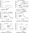
Assessment of barrier integrity in corneal endothelial monolayers. (A) Loss of barrier integrity shown by increased flux of HRP (horseradish peroxidase) across endothelial monolayer grown on Anopore™ filters. OD on the Y-axis represents optical density proportional to permeability. T: thrombin; C: Control; Y: Y-27632 (Rho kinase inhibitor). (B) Measurement of TER by electrical impedance across corneal endothelial monolayers grown on Anopore™ filters. Ado: Adenosine. (C) TER measurements using ECIS-1600R (Applied Biophysics, Inc., Troy, NY), including attachment and spreading of cells on the gold electrodes. After inoculation of the electrodes, impedance was measured at 3 frequencies at > 1 Hz. The plot shows the increase in the resistive component of the impedance at 4 KHz. A steady state is reached at about 18 hrs after cells grow to confluence. The resistance thus measured is a measure of the TER, which is taken to specify the barrier integrity quantitatively. (D) Loss of barrier integrity in response to cytochalasin D. After the normalized resistance had reached a steady state, the serum-rich medium in the electrode wells was replaced with a serum-free medium. At up-arrow shown at about 1 hr, cells were exposed to different levels of cytochalasin D, which is well known to break down the barrier integrity by depolymerization of actin cytoskeleton. As expected, cytochalasin D led to a decrease in the normalized resistance in a dose dependent manner. Summarized from our previous publications.–, , –
Figure 10
Figure 10
Breakdown and reassembly of apical junctions during Ca2+ Switch: (A) Disassembly of AJs by Ca2+ removal: Cells grown on gold electrodes were exposed to a Ca2+-free Ringers (w/ 2 mM EGTA). This led to an immediate precipitous fall in TER consistent with the breakdown of AJs. (B) Neoformation of AJs and TJs: Ca2+ add-back led to a gradual recovery of TER. The rate and extent of recovery was dependent on the level of extracellular Ca2+. (C) Disassembly of AJs is opposed by Y-27632 (Rho kinase inhibitor). Cells pretreated with Y-27632 were exposed Ca2+-free Ringers. The rate of decrease in TER was smaller compared to the rate observed in the absence of Y-27632. (D) Disassembly of AJs is opposed by blebbistatin (Myosin II ATPase inhibitor): Cells pretreated w/ blebbistatin for 10 min were exposed Ca2+-free Ringers. The rate of decrease in TER was smaller compared to the rate observed in the absence of blebbistatin. Data in Panels B–D, taken together, suggest a role for actomyosin contraction in the disassembly of AJs. Summarized from our previous publications.
Figure 11
Figure 11
Simplified overview of disassembly and reassembly of the AJC in the corneal endothelium during Ca2+ switch maneuver. We have shown that Ca2+ depletion results in disengagement of cadherins (i.e., breakdown of AJs) as well as outside-in signaling, resulting in activation of RhoA. Our results based on Y-27632 and blebbistatin further show that the consequent increase in actomyosin contraction of the PAMR accelerates breakdown of the TJs, which is induced by the disassembly of the AJs. Our Ca2+ add-back data have highlighted that F-actin is essential for the formation of AJs and that actomyosin contraction is crucial for the reassembly of TJs. Taken from our previous publication.
Figure 12
Figure 12
Effect of microtubule disassembly on MLC phosphporylation: (A) MLC phosphorylation in response to nocodazole (NDZ) is inhibited by Y-27632 (Rho kinase inhibitor; Y), C: Control (Untreated cells). (B) Bar graph of data similar to experiments shown in Panel A. (C) Nocodazole-mediated RhoA activation. One possible mechanism may involve release of GEF-H1 bound to microtubules. Taken from our previous publication.
Figure 13
Figure 13
Effect of paclitaxel on the (TNF-α)-induced response in corneal endothelial monolayers. (A) Effect of microtubule stabilization on TNF-α-induced microtubule disassembly. Cells were pretreated with 10 µM paclitaxel (PTX) for 1 hr with or without 20 ng/mL TNF-α for 6 hrs. Cells were then stained for α-tubulin (green) and counterstained for nucleus with DAPI (blue). In untreated cells, microtubules exist as fibrillary extensions from around the nucleus to the cell periphery (indicated by arrows). TNF-α induced microtubule disassembly is characterized by the loss as well as condensation of fibrillary extensions (indicated by arrows). Paclitaxel stabilize microtubule assembly and its pretreatment opposes microtubule disassembly by TNF-α. (B) Effect of microtubule stabilization on the (TNF-α)-induced dispersion of ZO-1. The influence of TNF-α on integrity of TJs was assessed by visualizing localization of ZO-1, a marker of TJ assembly, by immunostaining. Cells were pretreated with paclitaxel as in Panel A. ZO-1 at the plane of apical junctional complex is continuous throughout the cell periphery (shown by arrows) in untreated cells. TNF-α induces dispersion of ZO-1 from its normal locus. Upon treatment with paclitaxel, the pattern of ZO-1 localization is similar to those of untreated cells. (C) and (D): Effect of paclitaxel on TNF-α-induced decline in TER. Cells were pretreated with 10 µM paclitaxel for 1 hr with or without 20 ng/ml TNF-α. Paclitaxel attenuates the decrease in TER by TNF-α. Panel D shows bar graph of experiments similar to that shown in Panel C. The % reduction in TER induced by TNF-α is significantly greater than control beyond 8 hrs. Note: TNF-α + PTX: TNF-α +Paclitaxel. Error bars represent the SEM (n = 6). *Indicates significant difference from the control. C vs. TNF-α: p<0.001. Paclitaxel significantly opposes TNF-α-induced reduction in TER beyond 12 hrs. **Indicates significant difference TNF-α vs. TNF-α + PTX: p<0.001. Summarized from our previous publication.
Figure 14
Figure 14
p38 MAP kinase-dependent TNF-α response in corneal endothelial monolayers. (A) Effect of inhibition of p38 MAP kinase on TNF-α-induced microtubule disassembly. Cells were pretreated with 20 µM SB-203580 (SB) for 1 hr with or without 20 ng/mL TNF-α for 6 hrs. TNF-α induced microtubule disassembly is opposed by SB-203580 pre-treatment. (B) Effect of inhibition of p38 MAP kinase on the (TNF-α)-induced dispersion of ZO-1. Cells were pretreated with SB-203580 as in Panel A. Upon treatment with SB-203580, the pattern of ZO-1 localization is similar to those of untreated cells. (C) and (D): Effect of SB-203580 on TNF-α-induced decline in TER. Pretreatment with SB-203580 attenuates the decrease in TER by TNF-α. Panel D shows a bar graph of experiments similar to that shown in Panel C. **Significantly different than TNF-α, P < 0.001. Summarized from our previous publication.
Figure 15
Figure 15
Plasticity of the actin cytoskeleton. Extracellular stresses involving cytokines and neurohormonal stimuli mobilize the activity of small GTPases of Rho family and/or stress kinases (e.g., p38 MAP kinase) and, in turn, produce actin remodeling, actomyosin contraction, and/or microtubule disassembly. These changes enable remodeling of the apical junctional complex leading to changes in barrier integrity, cell-cell adhesion, and intercellular communication.

Similar articles

Cited by

References

    1. Edelhauser HF. The balance between corneal transparency and edema: the Proctor Lecture. Invest Ophthalmol Vis Sci. 2006;47:1754–1767. - PubMed
    1. Maurice DM. The location of the fluid pump in the cornea. J Physiol. 1972;221:43–54. - PMC - PubMed
    1. Dikstein S, Maurice DM. The metabolic basis to the fluid pump in the cornea. J Physiol. 1972;221:29–41. - PMC - PubMed
    1. Riley M. Pump and leak in regulation of fluid transport in rabbit cornea. Curr Eye Res. 1985;4:371–376. - PubMed
    1. Fischbarg J, Maurice DM. An update on corneal hydration control. Exp Eye Res. 2004;78:537–541. - PubMed

MeSH terms