Review
. 2010 Mar;6(3):146-56.
doi: 10.1038/nrrheum.2009.278. Epub 2010 Feb 9.

Sensors of the innate immune system: their link to rheumatic diseases

Affiliations
Review

Sensors of the innate immune system: their link to rheumatic diseases

Argyrios N Theofilopoulos et al. Nat Rev Rheumatol. 2010 Mar.

Abstract

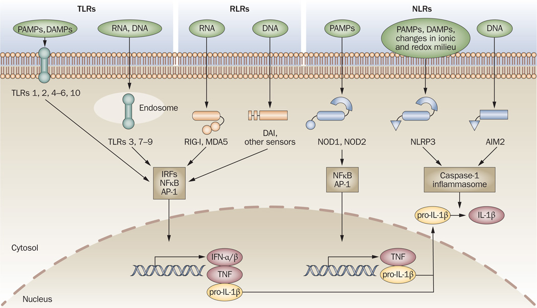
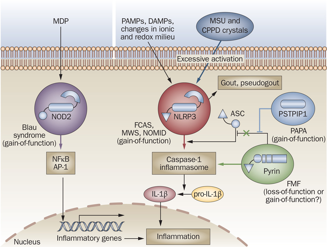
Evidence strongly suggests that excessive or protracted signaling, or both, by cell-surface or intracellular innate immune receptors is central to the pathogenesis of most autoimmune and autoinflammatory rheumatic diseases. The initiation of aberrant innate and adaptive immune responses in autoimmune diseases can be triggered by microbes and, at times, by endogenous molecules--particularly nucleic acids and related immune complexes--under sterile conditions. By contrast, most autoinflammatory syndromes are generally dependent on germline or de novo gene mutations that cause or facilitate inflammasome assembly. The consequent production of proinflammatory cytokines, principally interferon-alpha/beta and tumor necrosis factor in autoimmune diseases, and interleukin-1beta in autoinflammatory diseases, leads to the creation of autoamplification feedback loops and chronicity of these syndromes. These findings have resulted in a critical reappraisal of pathogenetic mechanisms, and provide a basis for the development of novel diagnostic and therapeutic modalities for these diseases.


Figures

Figure 1
Figure 1
Innate immune sensors. TLRs, RLRs (for example, RIG-I, MDA5, DAI and other sensors) and NLRs (for example, NODI, N0D2, NLRP3 and AIM2) are innate immune sensors that recognize danger signals derived from pathogens (PAMPs), damaged cells (DAMPs) or associated nucleic acids at the cell surface, in endolysosomes or in the cytoplasm. Signaling by these sensors promotes either the activation and nuclear translocation of transcription factors (IRFs, NFκB and AP-1) that drive expression of cytokines (IFN-α/β, TNF and pro-IL-lβ), or the assembly of the caspase-1 inflammasome and subsequent maturation of IL-1β from pro-IL-1β. Abbreviations: AIM2, absent in melanoma 2; AP-1, activator protein 1; DAMP, danger-associated molecular pattern; IFN, interferon; IL-1β, interleukin-1β; IRFs, interferon regulatory factors; MDA5, melanoma differentiation-associated gene-5; NFκB, nuclear factor κB; NLR, NOD-like receptor; NLRP, NLR with a pyrin domain; NOD, nucleotide-binding and oligomerization domain; PAMP, pathogen-associated molecular pattern; RIG-I, retinoid acid-inducible gene-l; RLR, RIG-l-like receptor; TLR, Toll-like receptor; TNF, tumor necrosis factor.
Figure 2
Figure 2
Breaching barriers for TLR recognition of self nucleic acids in systemic autoimmunity. Autoantibodies and autoreactive BCRs (for example, RFand anti-snRNP or anti-DNA antibodies) mediate the access of self nucleic acids to endolysosomal TLRs, leading to the production of type I IFNs from pDCs and autoantibodies of corresponding specificities from B cells. Accessory proteins such as HMGB1 (probably through RAGE) and LL37 can also facilitate DNA uptake and TLR engagement. The major snRNP antigen for anti-snRNP autoantibodies contains snRNA (red) and proteins (particle in yellow), whereas the antigenic targets of anti-DNA autoantibodies are apoptotic or necrotic materials containing DNA (blue), RNA (red) and other nucleic acid-binding molecules (particle in purple); accordingly, anti-RNP B cells are activated by TLR7 ligands, whereas anti-DNA or RF-producing B cells are activated by either TLR9 or TLR7 ligands. Abbreviations: BCR, B-cell receptor; FcγR, Feγ receptor; HMGB1, high mobility group box 1; IFN, interferon; pDC, plasmacytoid dendritic cell; RAGE, receptor for advanced glycosylation end-products; RF, rheumatoid factor; snRNA, small nuclear RNA; snRNP, small nuclear ribonucleoprotein; TLR, Toll-like receptor.
Figure 3
Figure 3
Trafficking of nucleic-acid-sensing TLRs. The ER-resident protein Unc93bl physically associates with TLR3, TLR7, TLR8 and TLR9 in the ER and mediates the trafficking of these receptors to endolysosomes. Efficient signaling by some of these TLRs might require proteolytic cleavage of a portion of the LRR ectodomain by lysosomal cathepsins and other proteases. Bacteria, viruses and other materials enter the cell through the endocytic pathway. Phagosomes and endosomes undergo interconnected maturation and fuse with lysosomes, giving rise to phagolysosomes or endolysosomes, acidic organelles that are rich in hydrolytic enzymes, in which degradation of the engulfed materials occurs. TLR engagement by the released nucleic acids leads to activation of transcription factors and expression of genes encoding cytokines. Abbreviations: ER, endoplasmic reticulum; LRR, leucine-rich repeat; TLR, Toll-like receptor.
Figure 4
Figure 4
Initiation and amplification stages in systemic autoimmunity. The pathogenesis of SLE might follow a two-phase process. In the first, initiation, phase (1), apoptotic or microbial materials, or both, induce TLR-independent or TLR-dependent production of type I IFNs by immature DCs, leading to DC maturation and efficient presentation of self-antigens, engagement of autoreactive T cells and B cells, and production of autoantibodies. In the second, self-sustaining amplification, phase (2), autoantibodies complexed with nucleic-acid-containing particles (RNP apoptotic or necrotic materials) are taken up by pDCs (via FcγR) and B cells (via BCR), leading to enhanced TLR-dependent type I IFN production, secretion of B-cell trophic factors, Ig class switching and perpetuation of the autoimmune response. Abbreviations: BCR, B-cell receptor; DC, dendritic cell; FcγR, Feγ receptor; IFN, interferon; pDC, plasmacytoid dendritic cell; RNP, ribonucleoprotein; SLE, systemic lupus erythematosus; TLR, Toll-like receptor.
Figure 5
Figure 5
Molecular basis of autoinflammatory diseases. Mutations in several components of the NLR signaling pathways promote uncontrollable spontaneous or ligand (MDP, PAMP, DAMP)-induced activation of either transcription factors (NFκB, AP-1) or the caspase-1 inflammasome, leading to excessive production of IL-1β and inflammation. Gene alterations identified so far result in gain-of-function mutations of NOD2 (in Blau syndrome), NLRP3 (in FCAS, MWS and NOMID) or PSTPIP1 (in PAPA), and probably loss-of-function mutations in pyrin (in FMF), which lead to inefficient sequestration of ASC, an adaptor required for inflammasome assembly. In addition, activation of NLRP3 by MSU and CPPD crystals is associated with the pathogenesis of gout and pseudogout, respectively. Abbreviations: AP-1, activator protein 1; ASC, apoptosis-associated speck-like protein containing a caspase activation and recruitment domain (CARD); CPPD, calcium pyrophosphate dihydrate; DAMP, danger-associated molecular pattern; FCAS, familial cold autoinflammatory syndrome; FMF, familial Mediterranean fever; IL-1β, interleukin-1β; MDP, muramyl dipeptide; MSU, monosodium urate; MWS, Muckle– Wells syndrome; NFκB, nuclear factor κB; NLR, NOD-like receptor; NLRR, NLR with a pyrin domain; NOD, nucleotide-binding and oligomerization domain; NOMID, neonatal onset multisystem inflammatory disease; PAPA, pyogenic arthritis with pyoderma gangrenosum and acne; PAMP, pathogen-associated molecular pattern; PSTPIP1, proline–serine–threonine–phosphatase-interacting protein 1.

Similar articles

Cited by

References

    1. Baccala R, et al. Sensors of the innate immune system: their mode of action. Nat. Rev. Rheumatol. 2009;5:448–456. - PubMed
    1. Ishii KJ, Akira S. TLR ignores methylated RNA? Immunity. 2005;23:111–113. - PubMed
    1. Schlee M, et al. Approaching the RNA ligand for RIG-I? Immunol. Rev. 2009;227:66–74. - PubMed
    1. Bave U, Vallin H, Alm GV, Ronnblom L. Activation of natural interferon-alpha producing cells by apoptotic U937 cells combined with Iupus IgG and its regulation by cytokines. J. Autoimmun. 2001;17:71–80. - PubMed
    1. Leadbetter EA, et al. Chromatin-IgG complexes activate B cells by dual engagement of IgM and Toll-like receptors. Nature. 2002;416:603–607. - PubMed

Publication types

MeSH terms