Fiber optic backscatter spectroscopic sensor to monitor enamel demineralization and remineralization in vitro
- PMID: 20142887
- PMCID: PMC2813093
- DOI: 10.4103/0972-0707.44053
Fiber optic backscatter spectroscopic sensor to monitor enamel demineralization and remineralization in vitro
Abstract
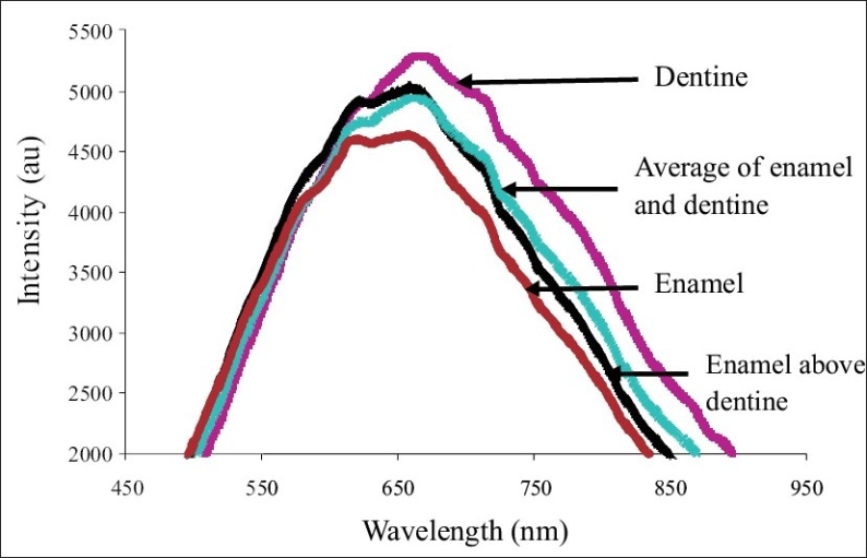
In this study, a Fiber Optic Backscatter Spectroscopic Sensor (FOBSS) is used to monitor demineralization and remineralization induced changes in the enamel. A bifurcated fiber optic backscatter probe connected to a visible light source and a high resolution spectrophotometer was used to acquire the backscatter light spectrum from the tooth surface. The experiments were conducted in two parts. In Part 1, experiments were carried out using fiber optic backscatter spectroscopy on (1) sound enamel and dentine sections and (2) sound tooth specimens subjected to demineralization and remineralization. In Part 2, polarization microscopy was conducted to examine the depth of demineralization in tooth specimens. The enamel and dentine specimens from the Part-1 experiments showed distinct backscatter spectra. The spectrum obtained from the enamel-dentine combination and the spectrum generated from the average of the enamel and dentine spectral values were closely similar and showed characteristics of dentine. The experiments in Part 2 showed that demineralization and remineralization processes induced a linear decrease and linear increase in the backscatter light intensity respectively. A negative correlation between the decrease in the backscatter light intensity during demineralization and the depth of demineralization determined using the polarization microscopy was calculated to be p = -0.994. This in vitro experiment highlights the potential benefit of using FOBSS to detect demineralization and remineralization of enamel.
Keywords: Fiber optic; demineralization; dentine; remineralization.
Conflict of interest statement
Figures


Similar articles
-
Demineralization of human dentine compared with enamel in a pH-cycling apparatus with a constant composition during de- and remineralization periods.Caries Res. 1991;25(5):317-22. doi: 10.1159/000261385. Caries Res. 1991. PMID: 1747881
-
In situ clinical effects of new dentifrices containing 1.5% arginine and fluoride on enamel de- and remineralization and plaque metabolism.J Clin Dent. 2013;24 Spec no A:A32-44. J Clin Dent. 2013. PMID: 24156138 Clinical Trial.
-
[Study on remineralization of human fluorosed teeth in vitro].Hua Xi Kou Qiang Yi Xue Za Zhi. 2009 Feb;27(1):96-9. Hua Xi Kou Qiang Yi Xue Za Zhi. 2009. PMID: 19323407 Chinese.
-
[Local enamel demineralization diagnostics and treatment].Stomatologiia (Mosk). 2017;96(4):67-71. doi: 10.17116/stomat201796467-71. Stomatologiia (Mosk). 2017. PMID: 28858285 Review. Russian.
-
High-resolution electron microscopy of enamel-crystal demineralization and remineralization in carious lesions.J Electron Microsc (Tokyo). 2003;52(6):605-13. doi: 10.1093/jmicro/52.6.605. J Electron Microsc (Tokyo). 2003. PMID: 14756249 Review.
Cited by
-
In vitro evaluation of the efficacy of laser fluorescence (DIAGNOdent) to detect demineralization and remineralization of smooth enamel lesions.J Conserv Dent. 2013 Jul;16(4):362-6. doi: 10.4103/0972-0707.114360. J Conserv Dent. 2013. PMID: 23956542 Free PMC article.
-
Morphological aspects in remineralizing potential of Silver Diamine Fluoride.Rom J Morphol Embryol. 2021 Apr-Jun;62(2):537-543. doi: 10.47162/RJME.62.2.20. Rom J Morphol Embryol. 2021. PMID: 35024742 Free PMC article.
References
-
- Stookey GK, Gonzalez-Cabezas C. Emerging methods of caries diagnosis. J Dent Educ. 2001;65:1001–6. - PubMed
-
- Stookey GK. The evolution of caries detection. Dimensions Dent Hyg. 2003;1:12–5.
-
- Abraham K. Lasers and optical fibers in medicine. St Louis, USA: Elsevier; 1993. pp. 111–38.
-
- Hintze H, Wenzle A, Danielsen B, Nyvad B. Reliability of visual examination, fibre-optics transillumination, bite-wing radiography and reproducibility of direct visual examination following tooth separation for the identification of cavitated carious lesions in contacting approximal surfaces. Caries Res. 1998;32:204–9. - PubMed
-
- Wenzel A, Larsen MJ, Fejerskov O. Detection of occlusal caries without cavitation by visual inspection, film radiographs, xeroradiographs, and digitized radiographs. Caries Res. 1991;25:365–71. - PubMed